University of Colorado-Denver, Dept. of Medicine/Cardiology, Aurora, CO, United States of America.
Medical Scientist Training Program, Aurora, CO, United States of America.
PLoS One. 2019 Aug 2;14(8):e0220573. doi: 10.1371/journal.pone.0220573. eCollection 2019.
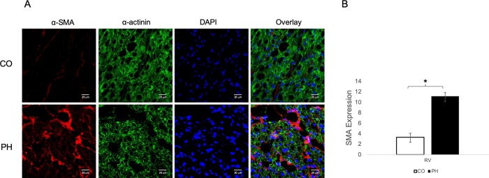
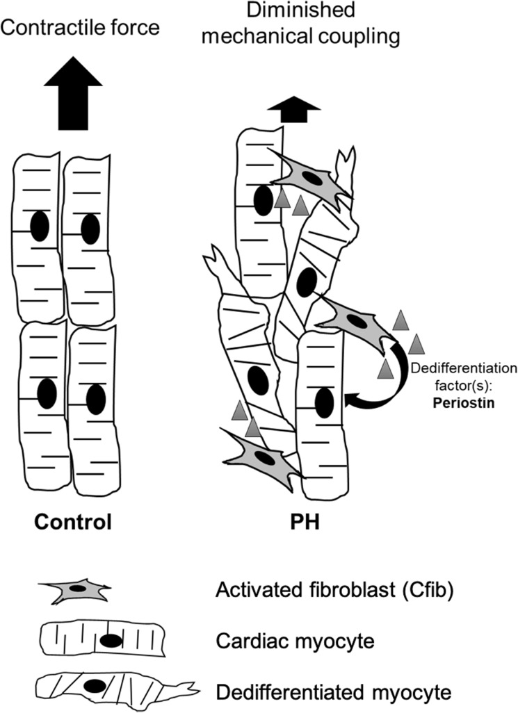
In virtually all models of heart failure, prognosis is determined by right ventricular (RV) function; thus, understanding the cellular mechanisms contributing to RV dysfunction is critical. Whole organ remodeling is associated with cell-specific changes, including cardiomyocyte dedifferentiation and activation of cardiac fibroblasts (Cfib) which in turn is linked to disorganization of cytoskeletal proteins and loss of sarcomeric structures. However, how these cellular changes contribute to RV function remains unknown. We've previously shown significant organ-level RV dysfunction in a large animal model of pulmonary hypertension (PH) which was not mirrored by reduced function of isolated cardiomyocytes. We hypothesized that factors produced by the endogenous Cfib contribute to global RV dysfunction by generating a heterogeneous cellular environment populated by dedifferentiated cells.
To determine the effect of Cfib conditioned media (CM) from the PH calf (PH-CM) on adult rat ventricular myocytes (ARVM) in culture.
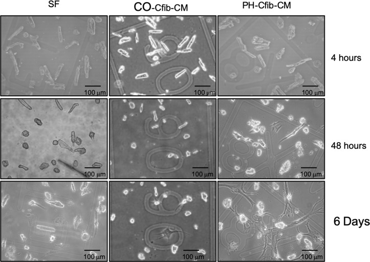
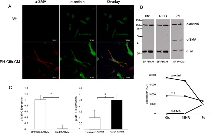
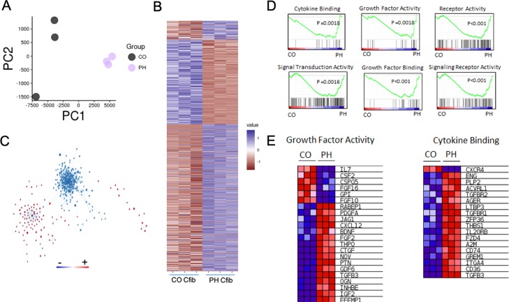
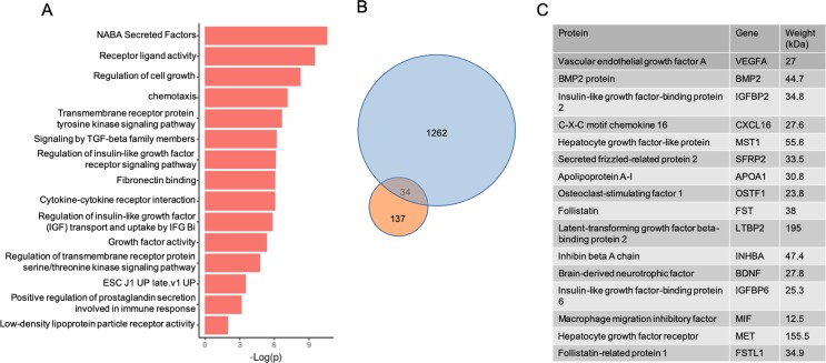
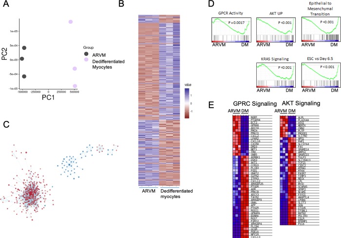
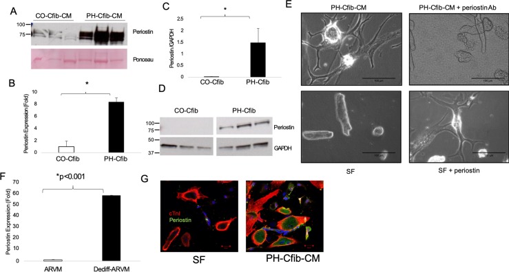
Brief exposure (<2 days) to PH-CM results in rapid, marked dedifferentiation of ARVM to a neonatal-like phenotype exhibiting spontaneous contractile behavior. Dedifferentiated cells maintain viability for over 30 days with continued expression of cardiomyocyte proteins including TnI and α-actinin yet exhibit myofibroblast characteristics including expression of α-smooth muscle actin. Using a bioinformatics approach to identify factor(s) that contribute to dedifferentiation, we found activation of the PH Cfib results in a unique transcriptome correlating with factors both in the secretome and with activated pathways in the dedifferentiated myocyte. Further, we identified upregulation of periostin in the Cfib and CM, and demonstrate that periostin is sufficient to drive cardiomyocyte dedifferentiation.
These data suggest that paracrine factor(s) released by Cfib from the PH calf signal a phenotypic transformation in a population of cardiomyocytes that likely contributes to RV dysfunction. Therapies targeting this process, such as inhibition of periostin, have the potential to prevent RV dysfunction.
几乎在所有心力衰竭模型中,预后都由右心室(RV)功能决定;因此,了解导致 RV 功能障碍的细胞机制至关重要。整个器官重塑与细胞特异性变化相关,包括心肌细胞去分化和心脏成纤维细胞(Cfib)的激活,这反过来又与细胞骨架蛋白的紊乱和肌节结构的丧失有关。然而,这些细胞变化如何导致 RV 功能仍然未知。我们之前在一种大型肺动脉高压(PH)动物模型中发现了显著的器官水平 RV 功能障碍,但与分离的心肌细胞功能降低不匹配。我们假设,内源性 Cfib 产生的因子通过产生由去分化细胞组成的异质细胞环境,导致整体 RV 功能障碍。
确定 PH 小牛(PH-CM)的 Cfib 条件培养基(CM)对培养中的成年大鼠心室肌细胞(ARVM)的影响。
短暂暴露(<2 天)于 PH-CM 可迅速、显著地使 ARVM 向类似于新生儿的表型去分化,表现出自发收缩行为。去分化细胞的存活率超过 30 天,同时持续表达肌钙蛋白 I 和α-辅肌动蛋白等心肌蛋白,但表现出肌成纤维细胞特征,包括表达α-平滑肌肌动蛋白。使用生物信息学方法来鉴定导致去分化的因子,我们发现 PH Cfib 的激活导致与分泌组中的因子以及去分化肌细胞中激活的途径相关的独特转录组。此外,我们发现 Cfib 和 CM 中的 periostin 上调,并证明 periostin 足以驱动心肌细胞去分化。
这些数据表明,PH 小牛的 Cfib 释放的旁分泌因子信号传递给一群心肌细胞,使它们发生表型转化,这可能导致 RV 功能障碍。针对该过程的治疗方法,如抑制 periostin,有可能预防 RV 功能障碍。