• 文献检索
  • 文档翻译
  • 深度研究
  • 学术资讯
  • Suppr Zotero 插件Zotero 插件
  • 邀请有礼
  • 套餐&价格
  • 历史记录
应用&插件
Suppr Zotero 插件Zotero 插件浏览器插件Mac 客户端Windows 客户端微信小程序
定价
高级版会员购买积分包购买API积分包
服务
文献检索文档翻译深度研究API 文档MCP 服务
关于我们
关于 Suppr公司介绍联系我们用户协议隐私条款
关注我们

Suppr 超能文献

核心技术专利:CN118964589B侵权必究
粤ICP备2023148730 号-1Suppr @ 2026

文献检索

告别复杂PubMed语法,用中文像聊天一样搜索,搜遍4000万医学文献。AI智能推荐,让科研检索更轻松。

立即免费搜索

文件翻译

保留排版,准确专业,支持PDF/Word/PPT等文件格式,支持 12+语言互译。

免费翻译文档

深度研究

AI帮你快速写综述,25分钟生成高质量综述,智能提取关键信息,辅助科研写作。

立即免费体验

过渡扩增细胞协调小鼠切牙间充质干细胞的激活。

Transit amplifying cells coordinate mouse incisor mesenchymal stem cell activation.

机构信息

Stem Cells & Regenerative Medicine Laboratory, Peninsula Dental School, University of Plymouth, 16 Research Way, Plymouth, PL6 8BU, UK.

Department of Cariology, Endodontology and Operative Dentistry, Peking University School and Hospital of Stomatology, 22 South Zhongguancun Avenue, Haidian District, 100081, Beijing, China.

出版信息

Nat Commun. 2019 Aug 9;10(1):3596. doi: 10.1038/s41467-019-11611-0.

DOI:10.1038/s41467-019-11611-0
PMID:31399601
原文链接:https://pmc.ncbi.nlm.nih.gov/articles/PMC6689115/
Abstract

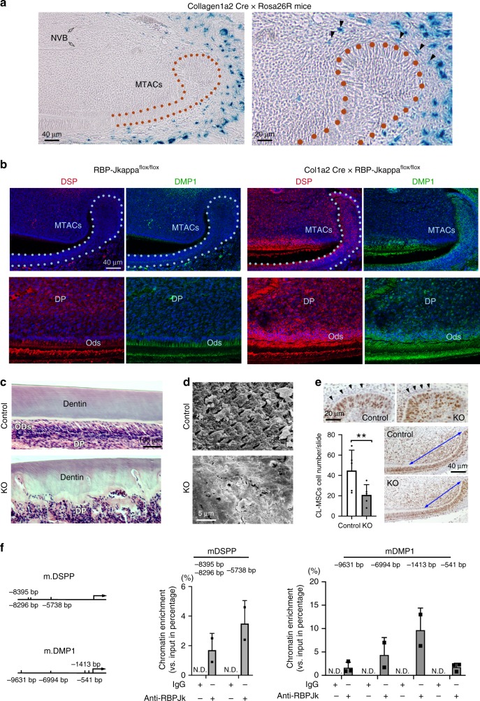
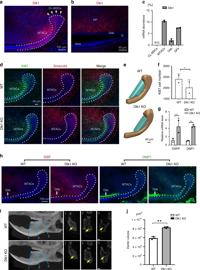
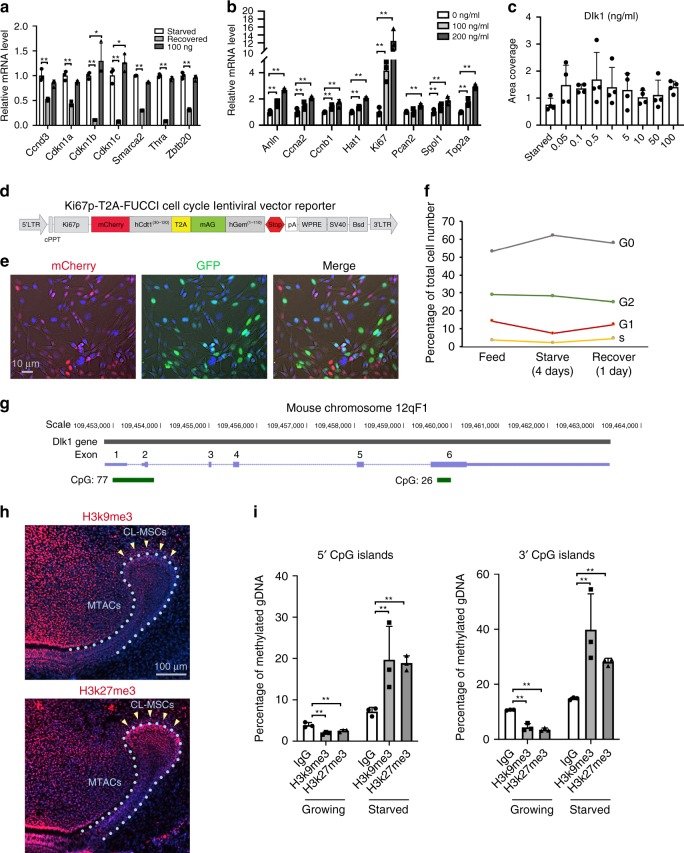
Stem cells (SCs) receive inductive cues from the surrounding microenvironment and cells. Limited molecular evidence has connected tissue-specific mesenchymal stem cells (MSCs) with mesenchymal transit amplifying cells (MTACs). Using mouse incisor as the model, we discover a population of MSCs neibouring to the MTACs and epithelial SCs. With Notch signaling as the key regulator, we disclose molecular proof and lineage tracing evidence showing the distinct MSCs contribute to incisor MTACs and the other mesenchymal cell lineages. MTACs can feedback and regulate the homeostasis and activation of CL-MSCs through Delta-like 1 homolog (Dlk1), which balances MSCs-MTACs number and the lineage differentiation. Dlk1's function on SCs priming and self-renewal depends on its biological forms and its gene expression is under dynamic epigenetic control. Our findings can be validated in clinical samples and applied to accelerate tooth wound healing, providing an intriguing insight of how to direct SCs towards tissue regeneration.

摘要

干细胞 (SCs) 从周围的微环境和细胞中接收诱导信号。有限的分子证据将组织特异性间充质干细胞 (MSCs) 与间充质过渡扩增细胞 (MTACs) 联系起来。我们使用小鼠切牙作为模型,发现了一群位于 MTACs 和上皮干细胞附近的 MSCs。以 Notch 信号作为关键调节因子,我们揭示了分子证据和谱系追踪证据,表明不同的 MSCs 有助于切牙 MTACs 和其他间充质细胞谱系。MTACs 可以通过 Delta-like 1 同源物 (Dlk1) 反馈和调节 CL-MSCs 的稳态和激活,从而平衡 MSCs-MTACs 的数量和谱系分化。Dlk1 在干细胞启动和自我更新中的功能取决于其生物形式,其基因表达受动态表观遗传控制。我们的发现可以在临床样本中得到验证,并应用于加速牙齿创伤愈合,为如何指导干细胞向组织再生提供了一个有趣的见解。

https://cdn.ncbi.nlm.nih.gov/pmc/blobs/23e7/6689115/b93acf170aed/41467_2019_11611_Fig9_HTML.jpg
https://cdn.ncbi.nlm.nih.gov/pmc/blobs/23e7/6689115/3c356f4f0d2e/41467_2019_11611_Fig1_HTML.jpg
https://cdn.ncbi.nlm.nih.gov/pmc/blobs/23e7/6689115/5db387878927/41467_2019_11611_Fig2_HTML.jpg
https://cdn.ncbi.nlm.nih.gov/pmc/blobs/23e7/6689115/2c552eed014f/41467_2019_11611_Fig3_HTML.jpg
https://cdn.ncbi.nlm.nih.gov/pmc/blobs/23e7/6689115/c0f382a0086e/41467_2019_11611_Fig4_HTML.jpg
https://cdn.ncbi.nlm.nih.gov/pmc/blobs/23e7/6689115/1a061686999e/41467_2019_11611_Fig5_HTML.jpg
https://cdn.ncbi.nlm.nih.gov/pmc/blobs/23e7/6689115/c89de0c44e9a/41467_2019_11611_Fig6_HTML.jpg
https://cdn.ncbi.nlm.nih.gov/pmc/blobs/23e7/6689115/a6740d62d152/41467_2019_11611_Fig7_HTML.jpg
https://cdn.ncbi.nlm.nih.gov/pmc/blobs/23e7/6689115/da327abc6a24/41467_2019_11611_Fig8_HTML.jpg
https://cdn.ncbi.nlm.nih.gov/pmc/blobs/23e7/6689115/b93acf170aed/41467_2019_11611_Fig9_HTML.jpg

相似文献

1
Transit amplifying cells coordinate mouse incisor mesenchymal stem cell activation.过渡扩增细胞协调小鼠切牙间充质干细胞的激活。
Nat Commun. 2019 Aug 9;10(1):3596. doi: 10.1038/s41467-019-11611-0.
2
Runx2+ Niche Cells Maintain Incisor Mesenchymal Tissue Homeostasis through IGF Signaling.Runx2+ 小生境细胞通过 IGF 信号维持切牙间充质组织稳态。
Cell Rep. 2020 Aug 11;32(6):108007. doi: 10.1016/j.celrep.2020.108007.
3
Ring1a/b polycomb proteins regulate the mesenchymal stem cell niche in continuously growing incisors.Ring1a/b 多梳蛋白调节不断生长的切牙中的间充质干细胞龛。
Dev Biol. 2012 Jul 15;367(2):140-53. doi: 10.1016/j.ydbio.2012.04.029. Epub 2012 May 4.
4
Regulation of Mesenchymal Stem to Transit-Amplifying Cell Transition in the Continuously Growing Mouse Incisor.调控间质干细胞向增殖活跃细胞转变在不断生长的小鼠切牙中
Cell Rep. 2018 Jun 5;23(10):3102-3111. doi: 10.1016/j.celrep.2018.05.001.
5
Reciprocal interaction between mesenchymal stem cells and transit amplifying cells regulates tissue homeostasis.间充质干细胞与过渡扩增细胞的相互作用调节组织内稳态。
Elife. 2021 Jan 22;10:e59459. doi: 10.7554/eLife.59459.
6
Secretion of shh by a neurovascular bundle niche supports mesenchymal stem cell homeostasis in the adult mouse incisor.神经血管束龛分泌的 shh 支持成年小鼠切牙间充质干细胞的体内平衡。
Cell Stem Cell. 2014 Feb 6;14(2):160-73. doi: 10.1016/j.stem.2013.12.013.
7
BMP Signaling in Regulating Mesenchymal Stem Cells in Incisor Homeostasis.BMP 信号在切牙稳态中调节间充质干细胞。
J Dent Res. 2019 Jul;98(8):904-911. doi: 10.1177/0022034519850812. Epub 2019 May 28.
8
Functionally Distinctive Ptch Receptors Establish Multimodal Hedgehog Signaling in the Tooth Epithelial Stem Cell Niche.功能不同的 Ptch 受体在牙齿上皮干细胞龛中建立多模式 Hedgehog 信号传导。
Stem Cells. 2019 Sep;37(9):1238-1248. doi: 10.1002/stem.3042. Epub 2019 Jun 10.
9
Negative effects of retinoic acid on stem cell niche of mouse incisor.视黄酸对小鼠切牙干细胞龛的负面影响。
Stem Cell Res. 2016 Nov;17(3):489-497. doi: 10.1016/j.scr.2016.09.030. Epub 2016 Sep 29.
10
Stromal DLK1 promotes proliferation and inhibits differentiation of the intestinal epithelium during development.基质细胞 DLK1 在肠道发育过程中促进肠道上皮细胞的增殖和抑制分化。
Am J Physiol Gastrointest Liver Physiol. 2021 Apr 1;320(4):G506-G520. doi: 10.1152/ajpgi.00445.2020. Epub 2021 Jan 20.

引用本文的文献

1
Din Oversees Mesenchymal Stem Cell Homeostasis in Mouse Incisors.Din调控小鼠切牙间充质干细胞的稳态。
Res Sq. 2025 May 14:rs.3.rs-6568233. doi: 10.21203/rs.3.rs-6568233/v1.
2
The Critical Balance Between Quiescence and Reactivation of Neural Stem Cells.神经干细胞静止与重新激活之间的关键平衡
Biomolecules. 2025 May 6;15(5):672. doi: 10.3390/biom15050672.
3
Glutamine-αKG axis affects dentin regeneration and regulates osteo/odontogenic differentiation of mesenchymal adult stem cells via IGF2 m6A modification.谷氨酰胺-α酮戊二酸轴通过IGF2 m6A修饰影响牙本质再生并调节间充质成体干细胞的骨/牙源性分化。

本文引用的文献

1
Prominin-1 controls stem cell activation by orchestrating ciliary dynamics.Prominin-1 通过协调纤毛动态控制干细胞激活。
EMBO J. 2019 Jan 15;38(2). doi: 10.15252/embj.201899845. Epub 2018 Dec 6.
2
Cre-driver lines used for genetic fate mapping of neural crest cells in the mouse: An overview.用于小鼠神经嵴细胞遗传命运图谱研究的Cre驱动系:综述。
Genesis. 2018 Jun;56(6-7):e23105. doi: 10.1002/dvg.23105. Epub 2018 Apr 19.
3
Soluble delta-like 1 homolog (DLK1) stimulates angiogenesis through Notch1/Akt/eNOS signaling in endothelial cells.
Stem Cell Res Ther. 2024 Dec 18;15(1):479. doi: 10.1186/s13287-024-04092-6.
4
Cathepsin K-Positive Cell Lineage Promotes In Situ Dentin Formation Controlled by Nociceptive Sonic Hedgehog.组织蛋白酶K阳性细胞谱系促进由伤害性音猬因子控制的原位牙本质形成。
Adv Sci (Weinh). 2024 Dec;11(47):e2310048. doi: 10.1002/advs.202310048. Epub 2024 Oct 30.
5
The Potential Reversible Transition between Stem Cells and Transient-Amplifying Cells: The Limbal Epithelial Stem Cell Perspective.干细胞与暂态扩增细胞之间的潜在可逆转化:角膜缘上皮干细胞视角。
Cells. 2024 Apr 25;13(9):748. doi: 10.3390/cells13090748.
6
Vascular architecture regulates mesenchymal stromal cell heterogeneity via P53-PDGF signaling in the mouse incisor.血管结构通过小鼠切牙中的 P53-PDGF 信号调节间充质基质细胞异质性。
Cell Stem Cell. 2024 Jun 6;31(6):904-920.e6. doi: 10.1016/j.stem.2024.04.011. Epub 2024 May 3.
7
Evaluation of the Efficacy of Stem Cells Therapy in the Periodontal Regeneration: A Meta-Analysis and Mendelian Randomization Study.评价干细胞疗法在牙周再生中的疗效:一项荟萃分析和孟德尔随机化研究。
Stem Cell Rev Rep. 2024 May;20(4):980-995. doi: 10.1007/s12015-024-10690-x. Epub 2024 Feb 22.
8
Dental Pulp Stem Cells and Current in vivo Approaches to Study Dental Pulp Stem Cells in Pulp Injury and Regeneration.牙髓干细胞及目前研究牙髓干细胞在牙髓损伤与再生中作用的体内研究方法
J Bone Metab. 2023 Aug;30(3):231-244. doi: 10.11005/jbm.2023.30.3.231. Epub 2023 Aug 31.
9
Novel Functional Peptide for Next-Generation Vital Pulp Therapy.用于下一代活髓治疗的新型功能肽。
J Dent Res. 2023 Mar;102(3):322-330. doi: 10.1177/00220345221135766. Epub 2022 Nov 22.
10
Deletion of RBP-Jkappa gene in mesenchymal cells causes rickets like symptoms in the mouse.间充质细胞中RBP-Jkappa基因的缺失会在小鼠中引发类似佝偻病的症状。
Curr Med (Cham). 2022;1(1):7. doi: 10.1007/s44194-022-00007-w. Epub 2022 May 26.
可溶性 delta 样配体 1 同源物(DLK1)通过内皮细胞中的 Notch1/Akt/eNOS 信号通路刺激血管生成。
Angiogenesis. 2018 May;21(2):299-312. doi: 10.1007/s10456-018-9596-7. Epub 2018 Jan 30.
4
A quiescent cell population replenishes mesenchymal stem cells to drive accelerated growth in mouse incisors.一个静止的细胞群体补充间充质干细胞以驱动小鼠门牙的加速生长。
Nat Commun. 2018 Jan 25;9(1):378. doi: 10.1038/s41467-017-02785-6.
5
Emerging roles of transit-amplifying cells in tissue regeneration and cancer.过渡放大细胞在组织再生和癌症中的新作用。
Wiley Interdiscip Rev Dev Biol. 2017 Sep;6(5). doi: 10.1002/wdev.282. Epub 2017 Jul 3.
6
Monitoring Notch Signaling-Associated Activation of Stem Cell Niches within Injured Dental Pulp.监测受损牙髓内干细胞龛的Notch信号相关激活
Front Physiol. 2017 May 30;8:372. doi: 10.3389/fphys.2017.00372. eCollection 2017.
7
Different expression levels of DLK1 inversely modulate the oncogenic potential of human MDA-MB-231 breast cancer cells through inhibition of NOTCH1 signaling.DLK1的不同表达水平通过抑制NOTCH1信号通路,反向调节人MDA-MB-231乳腺癌细胞的致癌潜能。
FASEB J. 2017 Aug;31(8):3484-3496. doi: 10.1096/fj.201601341RRR. Epub 2017 May 1.
8
An FAK-YAP-mTOR Signaling Axis Regulates Stem Cell-Based Tissue Renewal in Mice.一种黏着斑激酶-Yes相关蛋白-雷帕霉素靶蛋白信号轴调控小鼠基于干细胞的组织更新。
Cell Stem Cell. 2017 Jul 6;21(1):91-106.e6. doi: 10.1016/j.stem.2017.03.023. Epub 2017 Apr 27.
9
Local anesthetics induce autophagy in young permanent tooth pulp cells.局部麻醉剂可诱导年轻恒牙牙髓细胞发生自噬。
Cell Death Discov. 2015 Sep 7;1:15024. doi: 10.1038/cddiscovery.2015.24. eCollection 2015.
10
Endothelial cells are progenitors of cardiac pericytes and vascular smooth muscle cells.内皮细胞是心脏周细胞和血管平滑肌细胞的前体细胞。
Nat Commun. 2016 Aug 12;7:12422. doi: 10.1038/ncomms12422.