Mader Edward C, Ramos Alexander B, Cruz Roberto A, Branch Lionel A
1 Louisiana State University Health Sciences Center, New Orleans, LA, USA.
J Investig Med High Impact Case Rep. 2019 Jan-Dec;7:2324709619868266. doi: 10.1177/2324709619868266.
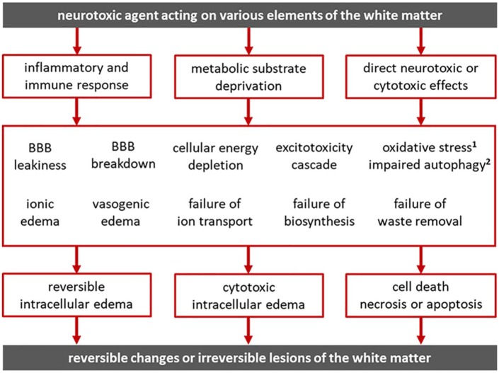
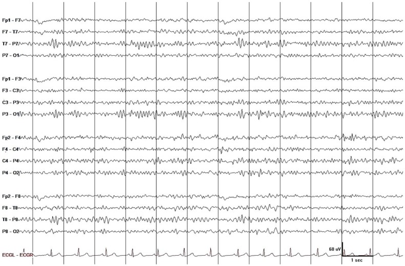
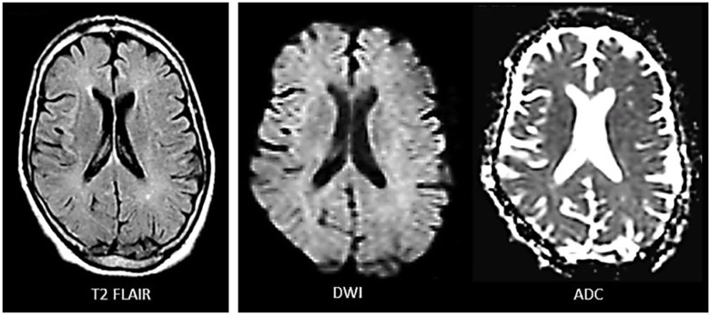
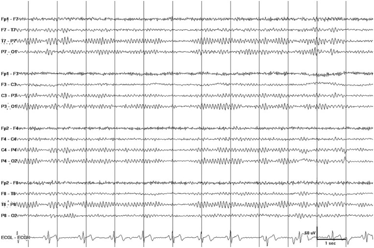
Toxic leukoencephalopathy (TL) is characterized by white matter disease on magnetic resonance imaging (MRI) and evidence of exposure to a neurotoxic agent. We describe a case of cocaine-induced TL in which extensive white matter disease did not preclude full recovery. A 57-year-old man with substance abuse disorder presented with a 5-day history of strange behavior. On admission, he was alert but had difficulty concentrating, psychomotor retardation, and diffuse hyperreflexia. Brain MRI revealed confluent subcortical white matter hyperintensities with restricted diffusion in some but not in other areas. Electroencephalography (EEG) showed mild diffuse slowing. Blood tests were normal except for mild hyperammonemia. Urine screen was positive for cocaine and benzodiazepine but quantitative analysis was significant only for cocaine. Prednisone 60-mg qd was initiated on day 4, tapered over a 5-day period, and discontinued on day 9. He was discharged after 3 weeks. Cognitive function returned to normal 2 weeks after discharge. Five months later, neurologic exam and EEG were normal and MRI showed near-100% resolution of white matter lesions. TL has been attributed to white matter ischemia/hypoxia resulting in demyelination/axonal injury. The clinical, EEG, and MRI findings and time course of recovery of our patient suggest that cocaine-induced inflammation/edema resulted in TL but not in ischemic/hypoxic injury. While inflammation/edema may have regressed when cocaine was discontinued, we cannot exclude a role for prednisone in protecting the patient from the ischemic/hypoxic sequelae of inflammation/edema. MRI is indispensable for diagnosing TL but EEG may provide additional useful diagnostic and prognostic information.
中毒性白质脑病(TL)的特征是磁共振成像(MRI)显示白质病变以及有接触神经毒性物质的证据。我们描述了一例可卡因诱发的TL病例,其中广泛的白质病变并未妨碍完全康复。一名患有物质使用障碍的57岁男性,有5天奇怪行为的病史。入院时,他神志清醒,但存在注意力不集中、精神运动迟缓及弥漫性反射亢进。脑部MRI显示皮质下白质融合性高信号,部分区域弥散受限,部分区域则无。脑电图(EEG)显示轻度弥漫性减慢。血液检查除轻度高氨血症外均正常。尿液筛查可卡因和苯二氮卓呈阳性,但定量分析仅可卡因含量显著。第4天开始使用泼尼松60mg每日一次,5天内逐渐减量,第9天停药。3周后他出院。出院后2周认知功能恢复正常。5个月后,神经系统检查和EEG正常,MRI显示白质病变几乎100%消退。TL被认为是白质缺血/缺氧导致脱髓鞘/轴索损伤。我们患者的临床、EEG、MRI表现及恢复时间过程提示,可卡因诱发的炎症/水肿导致了TL,但并非缺血/缺氧性损伤。虽然停用可卡因后炎症/水肿可能已消退,但我们不能排除泼尼松在保护患者免受炎症/水肿的缺血/缺氧后遗症方面的作用。MRI对诊断TL必不可少,但EEG可能提供额外有用的诊断和预后信息。