• 文献检索
  • 文档翻译
  • 深度研究
  • 学术资讯
  • Suppr Zotero 插件Zotero 插件
  • 邀请有礼
  • 套餐&价格
  • 历史记录
应用&插件
Suppr Zotero 插件Zotero 插件浏览器插件Mac 客户端Windows 客户端微信小程序
定价
高级版会员购买积分包购买API积分包
服务
文献检索文档翻译深度研究API 文档MCP 服务
关于我们
关于 Suppr公司介绍联系我们用户协议隐私条款
关注我们

Suppr 超能文献

核心技术专利:CN118964589B侵权必究
粤ICP备2023148730 号-1Suppr @ 2026

文献检索

告别复杂PubMed语法,用中文像聊天一样搜索,搜遍4000万医学文献。AI智能推荐,让科研检索更轻松。

立即免费搜索

文件翻译

保留排版,准确专业,支持PDF/Word/PPT等文件格式,支持 12+语言互译。

免费翻译文档

深度研究

AI帮你快速写综述,25分钟生成高质量综述,智能提取关键信息,辅助科研写作。

立即免费体验

多色成像在中心性浆液性脉络膜视网膜病变中的应用——与眼底自发荧光的定量和定性比较。

Multicolor imaging in central serous chorioretinopathy - a quantitative and qualitative comparison with fundus autofluorescence.

机构信息

Smt. Kanuri Santhamma Centre for Vitreo-Retinal Diseases, L V Prasad Eye Institute, Hyderabad, India.

Ophthalmology and Visual Sciences department, Khoo Teck Puat Hospital(KTPH), 90 Yishun Central, Singapore, Singapore.

出版信息

Sci Rep. 2019 Aug 13;9(1):11728. doi: 10.1038/s41598-019-48040-4.

DOI:10.1038/s41598-019-48040-4
PMID:31409843
原文链接:https://pmc.ncbi.nlm.nih.gov/articles/PMC6692404/
Abstract

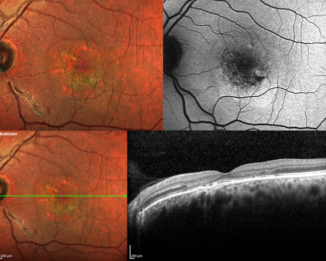
Central serous chorioretinopathy (CSCR) is characterised by choroidal hyperpermeability which results in neurosensory detachments (NSD) along with numerous retinal pigment epithelium (RPE) alterations such as RPE atrophy. Fundus autofluorescence (FAF) demonstrates the functionality of the RPE while multicolor imaging(MCI), by means of its three incident wavelengths, provides insight into clinical changes at various levels of the retina and choroid in CSCR. This study compares various clinical findings in CSCR (NSD, subretinal deposits, RPE atrophy, pigment epithelial detachments (PED) and pachyvessels) on the above mentioned imaging modalities both qualitatively and quantitatively. MCI showed higher mean cumulative area of RPE atrophic patches (6.3 ± 6.02 vs 5.7 ± 5.7 mm, p = 0.046), PED (1.3 ± 1.4 vs 1.1 ± 1.2 mm, p = 0.068) and NSD (17.2 ± 11.4 vs 15.7 ± 10.7 mm, p = 0.033). MCI demonstrated better defined lesions (NSD, PED, RPE atrophy) and more number of eyes with PED and pachyvessels in comparison to FAF.Both investigations had a 100% sensitivity in detecting NSD and 100% specificity for sub retinal deposits. This study demonstrates the ability of MCI to quantitatively and qualitatively define various clinical features in CSCR and the advantages it holds over FAF. MCI can hence be considered as a useful imaging modality in documenting and monitoring various structural changes in eyes with CSCR.

摘要

中心性浆液性脉络膜视网膜病变(CSCR)的特征是脉络膜通透性增加,导致神经感觉层脱离(NSD),同时伴有许多视网膜色素上皮(RPE)改变,如 RPE 萎缩。眼底自发荧光(FAF)显示 RPE 的功能,而多色成像(MCI)通过其三个入射波长,可以深入了解 CSCR 中视网膜和脉络膜各个层次的临床变化。本研究比较了 CSCR (NSD、视网膜下沉积物、RPE 萎缩、色素上皮脱离(PED)和厚血管)在上述成像方式下的各种临床发现,包括定性和定量两个方面。MCI 显示 RPE 萎缩斑块的平均累积面积(6.3±6.02 比 5.7±5.7mm,p=0.046)、PED(1.3±1.4 比 1.1±1.2mm,p=0.068)和 NSD(17.2±11.4 比 15.7±10.7mm,p=0.033)更大。与 FAF 相比,MCI 显示出更好定义的病变(NSD、PED、RPE 萎缩),并且更多的眼睛出现 PED 和厚血管。两种检查方法在检测 NSD 方面均具有 100%的敏感性,在检测视网膜下沉积物方面均具有 100%的特异性。本研究表明,MCI 能够定量和定性地定义 CSCR 中的各种临床特征,并且比 FAF 具有优势。因此,MCI 可以被认为是一种有用的成像方式,可用于记录和监测 CSCR 眼中的各种结构变化。

https://cdn.ncbi.nlm.nih.gov/pmc/blobs/e841/6692404/b2ebf51bfef9/41598_2019_48040_Fig3_HTML.jpg
https://cdn.ncbi.nlm.nih.gov/pmc/blobs/e841/6692404/a06dc238f919/41598_2019_48040_Fig1_HTML.jpg
https://cdn.ncbi.nlm.nih.gov/pmc/blobs/e841/6692404/83b102e01cf7/41598_2019_48040_Fig2_HTML.jpg
https://cdn.ncbi.nlm.nih.gov/pmc/blobs/e841/6692404/b2ebf51bfef9/41598_2019_48040_Fig3_HTML.jpg
https://cdn.ncbi.nlm.nih.gov/pmc/blobs/e841/6692404/a06dc238f919/41598_2019_48040_Fig1_HTML.jpg
https://cdn.ncbi.nlm.nih.gov/pmc/blobs/e841/6692404/83b102e01cf7/41598_2019_48040_Fig2_HTML.jpg
https://cdn.ncbi.nlm.nih.gov/pmc/blobs/e841/6692404/b2ebf51bfef9/41598_2019_48040_Fig3_HTML.jpg

相似文献

1
Multicolor imaging in central serous chorioretinopathy - a quantitative and qualitative comparison with fundus autofluorescence.多色成像在中心性浆液性脉络膜视网膜病变中的应用——与眼底自发荧光的定量和定性比较。
Sci Rep. 2019 Aug 13;9(1):11728. doi: 10.1038/s41598-019-48040-4.
2
Retro-mode Imaging for retinal pigment epithelium alterations in central serous chorioretinopathy.中心性浆液性脉络膜视网膜病变中视网膜色素上皮改变的反转模式成像。
Am J Ophthalmol. 2012 Jul;154(1):155-163.e4. doi: 10.1016/j.ajo.2012.01.023. Epub 2012 Apr 13.
3
Validation of multicolor imaging signatures of central serous chorioretinopathy lesions vis-a-vis conventional color fundus photographs.中心性浆液性脉络膜视网膜病变病变的多色成像特征相对于传统彩色眼底照片的验证。
Indian J Ophthalmol. 2020 May;68(5):861-866. doi: 10.4103/ijo.IJO_1187_19.
4
High-resolution adaptive optics-trans-scleral flood illumination (AO-TFI) imaging of retinal pigment epithelium (RPE) in central serous chorioretinopathy (CSCR).中心性浆液性脉络膜视网膜病变(CSCR)中视网膜色素上皮(RPE)的高分辨率自适应光学-经巩膜泛光照明(AO-TFI)成像。
Sci Rep. 2024 Jun 13;14(1):13689. doi: 10.1038/s41598-024-64524-4.
5
Scanning Laser Ophthalmoscopy Retromode Imaging Compared to Fundus Autofluorescence in Detecting Outer Retinal Features in Central Serous Chorioretinopathy.扫描激光眼底镜逆行成像与眼底自发荧光在检测中心性浆液性脉络膜视网膜病变外层视网膜特征方面的比较
Diagnostics (Basel). 2022 Oct 31;12(11):2638. doi: 10.3390/diagnostics12112638.
6
Multicolour imaging in central serous chorioretinopathy.多色成像在中心性浆液性脉络膜视网膜病变中的应用。
Clin Exp Optom. 2020 May;103(3):324-331. doi: 10.1111/cxo.12965. Epub 2019 Sep 6.
7
Retinal pigment epithelium apertures as a late complication of longstanding serous pigment epithelium detachments in chronic central serous chorioretinopathy.视网膜色素上皮孔是慢性中心性浆液性脉络膜视网膜病变中长期浆液性色素上皮脱离的晚期并发症。
Eye (Lond). 2019 Dec;33(12):1871-1876. doi: 10.1038/s41433-019-0505-0. Epub 2019 Jul 2.
8
Comparison of the optical coherence tomographic characters between acute Vogt-Koyanagi-Harada disease and acute central serous chorioretinopathy.急性 Vogt-小柳原田病与急性中心性浆液性脉络膜视网膜病变的光学相干断层扫描特征比较。
BMC Ophthalmol. 2014 Jun 28;14:87. doi: 10.1186/1471-2415-14-87.
9
Pathogenesis of Central Serous Chorioretinopathy and the Link Between Choroidal Hyperpermeability and Retinal Pigment Epithelium Pump Reversal.中心性浆液性脉络膜视网膜病变的发病机制及脉络膜高通透性与视网膜色素上皮泵功能逆转的关系。
Am J Ophthalmol. 2024 Oct;266:206-217. doi: 10.1016/j.ajo.2024.04.025. Epub 2024 May 7.
10
Fundus autofluorescence in acute and chronic central serous chorioretinopathy.急性和慢性中心性浆液性脉络膜视网膜病变中的眼底自发荧光
Clin Exp Optom. 2011 Sep;94(5):452-7. doi: 10.1111/j.1444-0938.2011.00598.x. Epub 2011 Apr 11.

引用本文的文献

1
Comparison of Multicolor Scanning Laser Imaging and Color Fundus Photography in Evaluating Vessel Whitening in Branch Retinal Vein Occlusion.多波长扫描激光成像与眼底彩色照相在评估分支型视网膜静脉阻塞中血管变白的比较。
Ophthalmic Res. 2023;66(1):413-420. doi: 10.1159/000528251. Epub 2022 Dec 8.
2
Association of Fundus Autofluorescence Abnormalities and Pachydrusen in Central Serous Chorioretinopathy and Polypoidal Choroidal Vasculopathy.中心性浆液性脉络膜视网膜病变和息肉状脉络膜血管病变中眼底自发荧光异常与玻璃膜疣的关联
J Clin Med. 2022 Sep 11;11(18):5340. doi: 10.3390/jcm11185340.
3
Commentary: Multicolor imaging: A new imaging tool in central serous chorioretinopathy.

本文引用的文献

1
Evolution of fundus autofluorescence patterns over time in patients with chronic central serous chorioretinopathy.慢性中心性浆液性脉络膜视网膜病变患者眼底自发荧光模式随时间的演变。
Acta Ophthalmol. 2018 Nov;96(7):e835-e839. doi: 10.1111/aos.13742. Epub 2018 Apr 15.
2
Comparison of conventional color fundus photography and multicolor imaging in choroidal or retinal lesions.传统彩色眼底照相术与多色成像在脉络膜或视网膜病变中的比较。
Graefes Arch Clin Exp Ophthalmol. 2018 Apr;256(4):643-649. doi: 10.1007/s00417-017-3884-6. Epub 2018 Feb 28.
3
IDENTIFYING FEATURES OF EARLY AND LATE AGE-RELATED MACULAR DEGENERATION: A Comparison of Multicolor Versus Traditional Color Fundus Photography.
述评:多色成像:中心性浆液性脉络膜视网膜病变的一种新型成像工具。
Indian J Ophthalmol. 2020 May;68(5):866-867. doi: 10.4103/ijo.IJO_2190_19.
4
Validation of multicolor imaging signatures of central serous chorioretinopathy lesions vis-a-vis conventional color fundus photographs.中心性浆液性脉络膜视网膜病变病变的多色成像特征相对于传统彩色眼底照片的验证。
Indian J Ophthalmol. 2020 May;68(5):861-866. doi: 10.4103/ijo.IJO_1187_19.
早期和晚期年龄相关性黄斑变性的特征识别:多光谱与传统眼底彩色照相比较。
Retina. 2018 Sep;38(9):1751-1758. doi: 10.1097/IAE.0000000000001777.
4
VISUALIZATION OF MACULAR PUCKER BY MULTICOLOR SCANNING LASER IMAGING.多波长扫描激光成像对黄斑皱襞的显示。
Retina. 2018 Feb;38(2):352-358. doi: 10.1097/IAE.0000000000001525.
5
Characteristics of Hyperautofluorescent Choroidal Vessels Using Fundus Camera Type Short-Wavelength Autofluorescence.使用短波长自发荧光眼底相机观察高自发荧光脉络膜血管的特征
Ophthalmologica. 2016;236(3):175-180. doi: 10.1159/000449262. Epub 2016 Sep 10.
6
Clinical Application of Multicolor Imaging Technology.多色成像技术的临床应用
Ophthalmologica. 2016;236(1):8-18. doi: 10.1159/000446857. Epub 2016 Jul 13.
7
Fundus autofluorescence imaging patterns in central serous chorioretinopathy according to chronicity.根据病程划分的中心性浆液性脉络膜视网膜病变的眼底自发荧光成像模式
Eye (Lond). 2016 Oct;30(10):1336-1342. doi: 10.1038/eye.2016.113. Epub 2016 Jun 10.
8
Optical Coherence Tomography Angiography in Central Serous Chorioretinopathy.中心性浆液性脉络膜视网膜病变的光学相干断层扫描血管造影
J Ophthalmol. 2015;2015:134783. doi: 10.1155/2015/134783. Epub 2015 Nov 8.
9
Central serous chorioretinopathy: Recent findings and new physiopathology hypothesis.中心性浆液性脉络膜视网膜病变:最新发现与新病理生理学假说。
Prog Retin Eye Res. 2015 Sep;48:82-118. doi: 10.1016/j.preteyeres.2015.05.003. Epub 2015 May 27.
10
Pachychoroid diseases of the macula.黄斑部厚脉络膜疾病
Med Hypothesis Discov Innov Ophthalmol. 2014 Winter;3(4):111-5.