Department of Orthopedics, Peking University First Hospital, Beijing 100034, P.R. China.
Mol Med Rep. 2019 Oct;20(4):3308-3316. doi: 10.3892/mmr.2019.10559. Epub 2019 Aug 6.
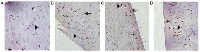
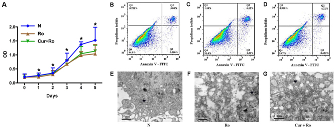
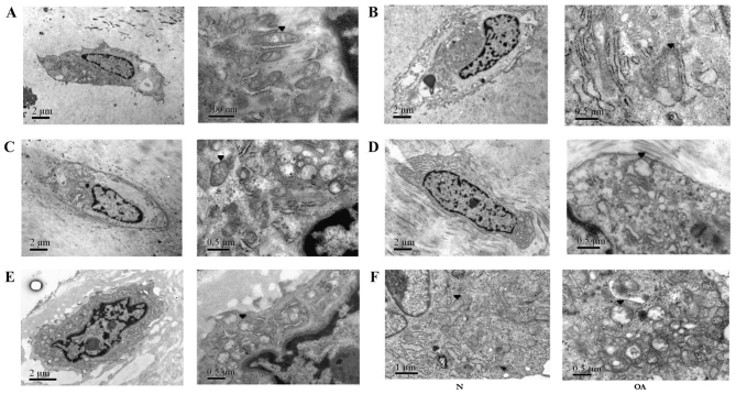
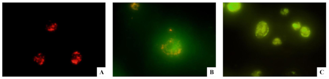
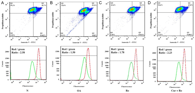
Previous studies identified that chondrocyte apoptosis serves an important role in osteoarthritis (OA). However, the mechanisms of cartilage degeneration induced by apoptosis remain unclear. The present study investigated the role of mitochondrial dysfunction in OA pathology. A total of 30 cartilage samples presenting an Outerbridge score ranging between 0 and III were collected during total knee arthroplasty. Half of the samples were embedded for observation by transmission electron microscopy. The remaining samples were digested, and chondrocytes were isolated from normal and OA tissues. Subsequently, the enzymatic activity of factors of the mitochondrial respiratory chain (MRC), and mitochondrial membrane potential (Δψm), were quantified. Furthermore, chondrocytes were treated with rotenone (Ro), a specific inhibitor of the MRC, and curcumin (Cur), a mitochondrial protective agent, with the aim of analyzing the relationship between mitochondrial dysfunction and chondrocyte apoptosis. The mitochondria of OA chondrocytes showed apoptosis‑associated morphological alterations compared with normal cells. The Δψm and the activity of MRC enzymes were decreased in OA chondrocytes. Moreover, compared with normal chondrocytes, treatment with Ro was able to induce morphological changes reminiscent of the phenotype observed in OA chondrocytes. Additionally, Ro inhibited cellular proliferation, increased the apoptotic rate, and decreased the Δψm and the secretion of type II collagen. Furthermore, Cur could partly reverse the effects caused by treatment with Ro. The present data suggested that mitochondrial function was impaired in OA chondrocytes, resulting in an increased chondrocyte apoptosis and decreased type II collagen secretion. In addition, treatment with Cur protected the mitochondrial function and prevented cartilage degeneration. Collectively, the present results suggested that mitochondrial dysfunction may aggravate cartilage degeneration in the pathogenesis of OA.
先前的研究表明,软骨细胞凋亡在骨关节炎(OA)中起着重要作用。然而,凋亡引起的软骨退化的机制尚不清楚。本研究探讨了线粒体功能障碍在 OA 病理中的作用。在全膝关节置换术中收集了 30 个软骨样本,这些样本的 Outerbridge 评分在 0 到 III 之间。将一半的样本包埋进行透射电子显微镜观察。其余样本进行消化,从正常和 OA 组织中分离软骨细胞。随后,定量测定线粒体呼吸链(MRC)因子和线粒体膜电位(Δψm)的酶活性。此外,用鱼藤酮(Ro),一种 MRC 的特异性抑制剂,和姜黄素(Cur),一种线粒体保护剂,处理软骨细胞,以分析线粒体功能障碍与软骨细胞凋亡之间的关系。与正常细胞相比,OA 软骨细胞的线粒体显示出与凋亡相关的形态改变。OA 软骨细胞中的 Δψm 和 MRC 酶的活性降低。此外,与正常软骨细胞相比,用 Ro 处理能够诱导类似于 OA 软骨细胞中观察到的表型的形态变化。此外,Ro 抑制细胞增殖,增加凋亡率,并降低 Δψm 和 II 型胶原的分泌。此外,Cur 可以部分逆转 Ro 处理引起的作用。这些数据表明,OA 软骨细胞中线粒体功能受损,导致软骨细胞凋亡增加和 II 型胶原分泌减少。此外,Cur 处理可保护线粒体功能并预防软骨退化。总之,这些结果表明线粒体功能障碍可能在 OA 的发病机制中加重软骨退化。