Department of Stereotactic and Functional Neurosurgery, Freiburg University Medical Center, Freiburg, Germany.
Medical Faculty, Freiburg University, Freiburg, Germany.
Transl Psychiatry. 2019 Aug 21;9(1):197. doi: 10.1038/s41398-019-0540-4.
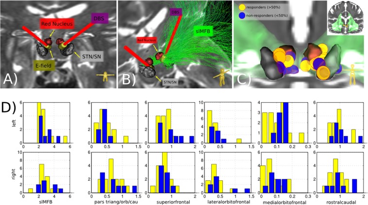
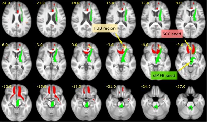
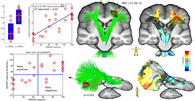
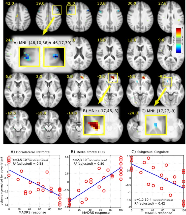
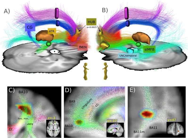
Major depression is a frequent and severe disorder, with a combination of psycho- and pharmacotherapy most patients can be treated. However, ~20% of all patients suffering from major depressive disorder remain treatment resistant; a subgroup might be treated with deep brain stimulation (DBS). We present two trials of DBS to the superolateral medial forebrain bundle (slMFB DBS; FORESEE I and II). The goal was to identify informed features that allow to predict treatment response. Data from N = 24 patients were analyzed. Preoperative imaging including anatomical sequences (T1 and T2) and diffusion tensor imaging (DTI) magnetic resonance imaging sequences were used together with postoperative helical CT scans (for DBS electrode position). Pathway activation modeling (PAM) as well as preoperative structural imaging and morphometry was used to understand the response behavior of patients (MADRS). A left fronto-polar and partly orbitofrontal region was identified that showed increased volume in preoperative anatomical scans. Further statistical analysis shows that the volume of this "HUB-region" is predictive for later MADRS response from DBS. The HUB region connects to typical fiber pathways that have been addressed before in therapeutic DBS in major depression. Left frontal volume growth might indicate intrinsic activity upon disconnection form the main emotional network. The results are significant since for the first time we found an informed feature that might allow to identify and phenotype future responders for slMFB DBS. This is a clear step into the direction of personalized treatments.
重度抑郁症是一种常见且严重的疾病,大多数患者可以通过心理和药物治疗相结合得到治疗。然而,约 20%的重度抑郁症患者仍然对治疗有抵抗,其中一部分患者可能会接受深部脑刺激(DBS)治疗。我们报告了两个对外侧眶额束(slMFB DBS;FORESEE I 和 II)进行 DBS 的试验。目的是确定有助于预测治疗反应的特征。对 n=24 名患者的数据进行了分析。术前影像包括解剖序列(T1 和 T2)和弥散张量成像(DTI)磁共振成像序列,以及术后螺旋 CT 扫描(用于 DBS 电极位置)。采用通路激活建模(PAM)以及术前结构成像和形态测量学来理解患者的反应行为(MADRS)。确定了左侧额极和部分眶额区域,在术前解剖扫描中显示出体积增加。进一步的统计分析表明,该“HUB 区域”的体积是 DBS 后 MADRS 反应的预测因素。HUB 区域连接到典型的纤维通路,这些通路在治疗性 DBS 治疗重度抑郁症中已经被研究过。左侧额叶体积的增长可能表明在与主要情感网络断开连接时的内在活动。这些结果意义重大,因为我们首次发现了一个有信息特征的特征,该特征可能有助于识别和表型未来对 slMFB DBS 的反应者。这是朝着个性化治疗迈出的明确一步。