• 文献检索
  • 文档翻译
  • 深度研究
  • 学术资讯
  • Suppr Zotero 插件Zotero 插件
  • 邀请有礼
  • 套餐&价格
  • 历史记录
应用&插件
Suppr Zotero 插件Zotero 插件浏览器插件Mac 客户端Windows 客户端微信小程序
定价
高级版会员购买积分包购买API积分包
服务
文献检索文档翻译深度研究API 文档MCP 服务
关于我们
关于 Suppr公司介绍联系我们用户协议隐私条款
关注我们

Suppr 超能文献

核心技术专利:CN118964589B侵权必究
粤ICP备2023148730 号-1Suppr @ 2026

文献检索

告别复杂PubMed语法,用中文像聊天一样搜索,搜遍4000万医学文献。AI智能推荐,让科研检索更轻松。

立即免费搜索

文件翻译

保留排版,准确专业,支持PDF/Word/PPT等文件格式,支持 12+语言互译。

免费翻译文档

深度研究

AI帮你快速写综述,25分钟生成高质量综述,智能提取关键信息,辅助科研写作。

立即免费体验

用于 FRET 生物传感器晶格光片成像的软件,用一个新的 Rap1 生物传感器进行说明。

Software for lattice light-sheet imaging of FRET biosensors, illustrated with a new Rap1 biosensor.

机构信息

Department of Pharmacology, University of North Carolina at Chapel Hill, Chapel Hill, NC.

Department of Biomedical Engineering, Georgia Institute of Technology and Emory University, Atlanta, GA.

出版信息

J Cell Biol. 2019 Sep 2;218(9):3153-3160. doi: 10.1083/jcb.201903019. Epub 2019 Aug 23.

DOI:10.1083/jcb.201903019
PMID:31444239
原文链接:https://pmc.ncbi.nlm.nih.gov/articles/PMC6719445/
Abstract

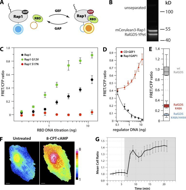
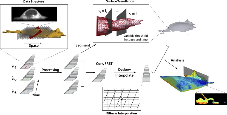
Lattice light-sheet microscopy (LLSM) is valuable for its combination of reduced photobleaching and outstanding spatiotemporal resolution in 3D. Using LLSM to image biosensors in living cells could provide unprecedented visualization of rapid, localized changes in protein conformation or posttranslational modification. However, computational manipulations required for biosensor imaging with LLSM are challenging for many software packages. The calculations require processing large amounts of data even for simple changes such as reorientation of cell renderings or testing the effects of user-selectable settings, and lattice imaging poses unique challenges in thresholding and ratio imaging. We describe here a new software package, named ImageTank, that is specifically designed for practical imaging of biosensors using LLSM. To demonstrate its capabilities, we use a new biosensor to study the rapid 3D dynamics of the small GTPase Rap1 in vesicles and cell protrusions.

摘要

晶格层光片显微镜(LLSM)在减少光漂白和在 3D 中具有出色的时空分辨率方面具有价值。在活细胞中使用 LLSM 对生物传感器进行成像,可以提供对蛋白质构象或翻译后修饰快速、局部变化的前所未有的可视化。然而,对于许多软件包来说,使用 LLSM 对生物传感器进行成像所需的计算操作具有挑战性。即使对于重新定向细胞渲染或测试用户可选设置的效果等简单变化,计算也需要处理大量数据,而晶格成像在阈值处理和比率成像方面存在独特的挑战。我们在这里描述了一个名为 ImageTank 的新软件包,它专门用于使用 LLSM 对生物传感器进行实际成像。为了展示其功能,我们使用一种新的生物传感器来研究小 GTPase Rap1 在小泡和细胞突起中的快速 3D 动力学。

https://cdn.ncbi.nlm.nih.gov/pmc/blobs/9d41/6719445/f2171edb757e/JCB_201903019_Fig4.jpg
https://cdn.ncbi.nlm.nih.gov/pmc/blobs/9d41/6719445/45bece564618/JCB_201903019_Fig1.jpg
https://cdn.ncbi.nlm.nih.gov/pmc/blobs/9d41/6719445/c185ae786a2a/JCB_201903019_Fig2.jpg
https://cdn.ncbi.nlm.nih.gov/pmc/blobs/9d41/6719445/bfa847ff0dbb/JCB_201903019_Fig3.jpg
https://cdn.ncbi.nlm.nih.gov/pmc/blobs/9d41/6719445/f2171edb757e/JCB_201903019_Fig4.jpg

相似文献

1
Software for lattice light-sheet imaging of FRET biosensors, illustrated with a new Rap1 biosensor.用于 FRET 生物传感器晶格光片成像的软件,用一个新的 Rap1 生物传感器进行说明。
J Cell Biol. 2019 Sep 2;218(9):3153-3160. doi: 10.1083/jcb.201903019. Epub 2019 Aug 23.
2
FRET Microscopy for Real-Time Visualization of Second Messengers in Living Cells.用于实时可视化活细胞中第二信使的荧光共振能量转移显微镜技术
Methods Mol Biol. 2017;1563:85-90. doi: 10.1007/978-1-4939-6810-7_6.
3
Rapid high resolution 3D imaging of expanded biological specimens with lattice light sheet microscopy.晶格层光片显微镜快速高分辨率 3D 成像扩展生物样本。
Methods. 2020 Mar 1;174:11-19. doi: 10.1016/j.ymeth.2019.04.006. Epub 2019 Apr 9.
4
In Vivo Quantification of Intramolecular FRET Using RacFRET Biosensors.使用RacFRET生物传感器对分子内荧光共振能量转移进行体内定量分析。
Methods Mol Biol. 2019;2040:275-297. doi: 10.1007/978-1-4939-9686-5_13.
5
SH2 Domain-Based FRET Biosensor for Measuring BCR-ABL Activity in Living CML Cells.用于测量活的慢性粒细胞白血病(CML)细胞中BCR-ABL活性的基于SH2结构域的荧光共振能量转移(FRET)生物传感器。
Methods Mol Biol. 2017;1555:513-534. doi: 10.1007/978-1-4939-6762-9_30.
6
Quantitative Imaging of FRET-Based Biosensors for Cell- and Organelle-Specific Analyses in Plants.用于植物细胞和细胞器特异性分析的基于荧光共振能量转移(FRET)的生物传感器的定量成像
Microsc Microanal. 2016 Apr;22(2):300-10. doi: 10.1017/S143192761600012X. Epub 2016 Feb 16.
7
LiveLattice: Real-time visualization of tilted light-sheet microscopy data using a memory-efficient transformation algorithm.LiveLattice:使用内存高效变换算法对倾斜光片显微镜数据进行实时可视化
bioRxiv. 2024 May 30:2024.05.28.596280. doi: 10.1101/2024.05.28.596280.
8
Application of FRET-Based Biosensor "ATeam" for Visualization of ATP Levels in the Mitochondrial Matrix of Living Mammalian Cells.基于荧光共振能量转移(FRET)的生物传感器“ATeam”在活的哺乳动物细胞线粒体基质中ATP水平可视化中的应用。
Methods Mol Biol. 2017;1567:231-243. doi: 10.1007/978-1-4939-6824-4_14.
9
Large Scale Bacterial Colony Screening of Diversified FRET Biosensors.多样化荧光共振能量转移生物传感器的大规模细菌菌落筛选
PLoS One. 2015 Jun 10;10(6):e0119860. doi: 10.1371/journal.pone.0119860. eCollection 2015.
10
Detection and Quantification of Intracellular Signaling Using FRET-Based Biosensors and High Content Imaging.使用基于荧光共振能量转移(FRET)的生物传感器和高内涵成像技术检测和定量细胞内信号传导
Methods Mol Biol. 2015;1335:131-61. doi: 10.1007/978-1-4939-2914-6_10.

引用本文的文献

1
TRIM9 Controls Growth Cone Responses to Netrin Through DCC and UNC5C.TRIM9通过DCC和UNC5C调控生长锥对Netrin的反应。
J Neurochem. 2025 Jan;169(1):e70002. doi: 10.1111/jnc.70002.
2
Molecular Spies in Action: Genetically Encoded Fluorescent Biosensors Light up Cellular Signals.分子间谍大显身手:基因编码荧光生物传感器点亮细胞信号。
Chem Rev. 2024 Nov 27;124(22):12573-12660. doi: 10.1021/acs.chemrev.4c00293. Epub 2024 Nov 13.
3
4D intravital imaging studies identify platelets as the predominant cellular procoagulant surface in a mouse hemostasis model.

本文引用的文献

1
BAK/BAX macropores facilitate mitochondrial herniation and mtDNA efflux during apoptosis.BAK/BAX 大孔促进线粒体突出和 mtDNA 外排凋亡过程中。
Science. 2018 Feb 23;359(6378). doi: 10.1126/science.aao6047.
2
Phase separation drives heterochromatin domain formation.相分离驱动异染色质结构域的形成。
Nature. 2017 Jul 13;547(7662):241-245. doi: 10.1038/nature22989. Epub 2017 Jun 21.
3
Applying systems-level spectral imaging and analysis to reveal the organelle interactome.应用系统级光谱成像和分析来揭示细胞器相互作用组。
4D 活体成像研究在小鼠止血模型中确定血小板为主要的细胞促凝表面。
Blood. 2024 Sep 5;144(10):1116-1126. doi: 10.1182/blood.2023022608.
4
pHusion - a robust and versatile toolset for automated detection and analysis of exocytosis.pHusion——用于自动化检测和分析胞吐作用的强大且通用的工具集。
J Cell Sci. 2024 Oct 15;137(20). doi: 10.1242/jcs.261828. Epub 2024 Jun 7.
5
Unravelling molecular dynamics in living cells: Fluorescent protein biosensors for cell biology.解析活细胞中的分子动力学:用于细胞生物学的荧光蛋白生物传感器
J Microsc. 2025 May;298(2):123-184. doi: 10.1111/jmi.13270. Epub 2024 Feb 15.
6
Computationally designed sensors detect endogenous Ras activity and signaling effectors at subcellular resolution.通过计算设计的传感器可在亚细胞分辨率下检测内源性Ras活性和信号效应器。
Nat Biotechnol. 2024 Dec;42(12):1888-1898. doi: 10.1038/s41587-023-02107-w. Epub 2024 Jan 25.
7
Cellular harmonics for the morphology-invariant analysis of molecular organization at the cell surface.用于细胞表面分子组织形态不变分析的细胞谐波。
Nat Comput Sci. 2023 Sep;3(9):777-788. doi: 10.1038/s43588-023-00512-4. Epub 2023 Sep 14.
8
Surface-guided computing to analyze subcellular morphology and membrane-associated signals in 3D.用于在三维空间中分析亚细胞形态和膜相关信号的表面引导计算。
bioRxiv. 2023 Apr 20:2023.04.12.536640. doi: 10.1101/2023.04.12.536640.
9
Surface-guided computing to analyze subcellular morphology and membrane-associated signals in 3D.用于分析三维亚细胞形态和膜相关信号的表面引导计算
ArXiv. 2023 Apr 12:arXiv:2304.06176v1.
10
Advances in NK cell therapy for brain tumors.脑肿瘤自然杀伤细胞疗法的进展。
NPJ Precis Oncol. 2023 Feb 15;7(1):17. doi: 10.1038/s41698-023-00356-1.
Nature. 2017 Jun 1;546(7656):162-167. doi: 10.1038/nature22369. Epub 2017 May 24.
4
A High-Content Assay for Biosensor Validation and for Examining Stimuli that Affect Biosensor Activity.一种用于生物传感器验证和检测影响生物传感器活性刺激因素的高内涵分析方法。
Curr Protoc Cell Biol. 2014 Dec 1;65:14.15.1-31. doi: 10.1002/0471143030.cb1415s65.
5
Lattice light-sheet microscopy: imaging molecules to embryos at high spatiotemporal resolution.晶格层光片显微镜:以高时空分辨率对分子和胚胎进行成像。
Science. 2014 Oct 24;346(6208):1257998. doi: 10.1126/science.1257998. Epub 2014 Oct 23.
6
Chemical tools for studying directed cell migration.用于研究定向细胞迁移的化学工具。
ACS Chem Biol. 2011 Nov 18;6(11):1164-74. doi: 10.1021/cb200299k. Epub 2011 Oct 20.
7
High cleavage efficiency of a 2A peptide derived from porcine teschovirus-1 in human cell lines, zebrafish and mice.猪捷申病毒 2A 肽在人细胞系、斑马鱼和小鼠中的高效切割效率。
PLoS One. 2011;6(4):e18556. doi: 10.1371/journal.pone.0018556. Epub 2011 Apr 29.
8
Reporting from the field: genetically encoded fluorescent reporters uncover signaling dynamics in living biological systems.现场报道:基因编码荧光报告蛋白揭示活体生物系统中的信号动态。
Annu Rev Biochem. 2011;80:375-401. doi: 10.1146/annurev-biochem-060409-093259.
9
An improved cerulean fluorescent protein with enhanced brightness and reduced reversible photoswitching.一种亮度增强、可逆光开关减少的改良天青荧光蛋白。
PLoS One. 2011 Mar 29;6(3):e17896. doi: 10.1371/journal.pone.0017896.
10
Biosensors for characterizing the dynamics of rho family GTPases in living cells.用于表征活细胞中rho家族GTP酶动力学的生物传感器。
Curr Protoc Cell Biol. 2010 Mar;Chapter 14:Unit 14.11.1-26. doi: 10.1002/0471143030.cb1411s46.