Division of Molecular Pathology, National Cancer Center Research Institute, Tokyo, Japan.
Department of Analytical Pathology, National Cancer Center Research Institute, Tokyo, Japan.
Cancer Sci. 2020 Aug;111(8):3057-3070. doi: 10.1111/cas.14514. Epub 2020 Jul 9.
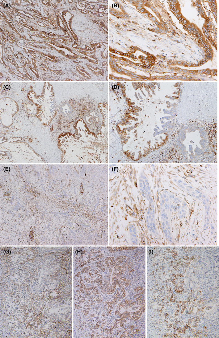
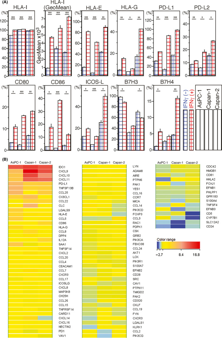
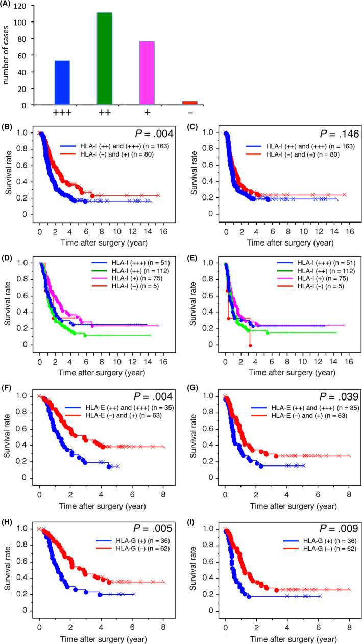
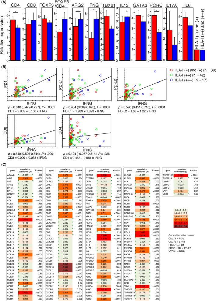
The expression of classical human leukocyte antigen class I antigens (HLA-I) on the surfaces of cancer cells allows cytotoxic T cells to recognize and eliminate these cells. Reduction or loss of HLA-I is a mechanism of escape from antitumor immunity. The present study aimed to investigate the clinicopathological impacts of HLA-I and non-classical HLA-I antigens expressed on pancreatic ductal adenocarcinoma (PDAC) cells. We performed immunohistochemistry to detect expression of HLA-I antigens in PDAC using 243 PDAC cases and examined their clinicopathological influences. We also investigated the expression of immune-related genes to characterize PDAC tumor microenvironments. Lower expression of HLA-I, found in 33% of PDAC cases, was significantly associated with longer overall survival. Higher expression of both HLA-E and HLA-G was significantly associated with shorter survival. Multivariate analyses revealed that higher expression of these three HLA-I antigens was significantly correlated with shorter survival. Higher HLA-I expression on PDAC cells was significantly correlated with higher expression of IFNG, which also correlated with PD1, PD-L1 and PD-L2 expression. In vitro assay revealed that interferon gamma (IFNγ) stimulation increased surface expression of HLA-I in three PDAC cell lines. It also upregulated surface expression of HLA-E, HLA-G and immune checkpoint molecules, including PD-L1 and PD-L2. These results suggest that the higher expression of HLA-I, HLA-E and HLA-G on PDAC cells is an unfavorable prognosticator. It is possible that IFNγ promotes a tolerant microenvironment by inducing immune checkpoint molecules in PDAC tissues with higher HLA-I expression on PDAC cells.
癌细胞表面经典人类白细胞抗原 I 类抗原(HLA-I)的表达使细胞毒性 T 细胞能够识别和消除这些细胞。HLA-I 的减少或丢失是逃避抗肿瘤免疫的一种机制。本研究旨在探讨 HLA-I 和非经典 HLA-I 抗原在胰腺导管腺癌(PDAC)细胞上的表达对临床病理的影响。我们使用 243 例 PDAC 病例进行免疫组织化学检测 HLA-I 抗原的表达,并检查其临床病理影响。我们还研究了免疫相关基因的表达,以描绘 PDAC 肿瘤微环境。在 33%的 PDAC 病例中发现 HLA-I 表达降低,与总生存时间延长显著相关。HLA-E 和 HLA-G 的表达较高与生存时间较短显著相关。多变量分析显示,这三种 HLA-I 抗原的高表达与较短的生存时间显著相关。PDAC 细胞上 HLA-I 的高表达与 IFNG 的高表达显著相关,IFNG 也与 PD1、PD-L1 和 PD-L2 的表达相关。体外实验表明,干扰素γ(IFNγ)刺激可增加三种 PDAC 细胞系表面 HLA-I 的表达。它还上调了 HLA-E、HLA-G 和免疫检查点分子的表面表达,包括 PD-L1 和 PD-L2。这些结果表明,PDAC 细胞上 HLA-I、HLA-E 和 HLA-G 的高表达是一个不利的预后因素。IFNγ 可能通过在 HLA-I 表达较高的 PDAC 细胞上诱导免疫检查点分子,在 HLA-I 表达较高的 PDAC 组织中促进耐受微环境。