Department of Radiation Physics, The University of Texas MD Anderson Cancer Center, Houston, Texas, United States of America.
MD Anderson Cancer Center UTHealth Graduate School of Biomedical Sciences, Houston, Texas, United States of America.
PLoS One. 2019 Sep 5;14(9):e0221877. doi: 10.1371/journal.pone.0221877. eCollection 2019.
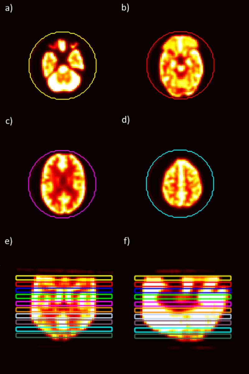
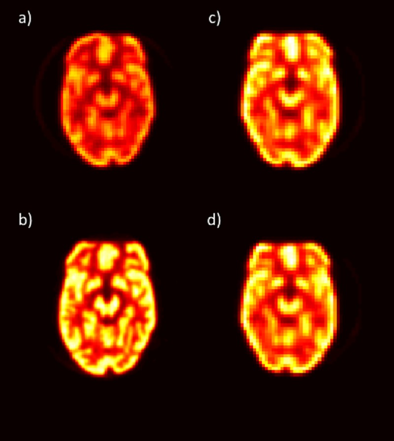
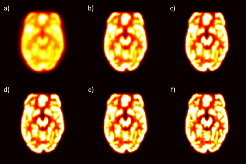
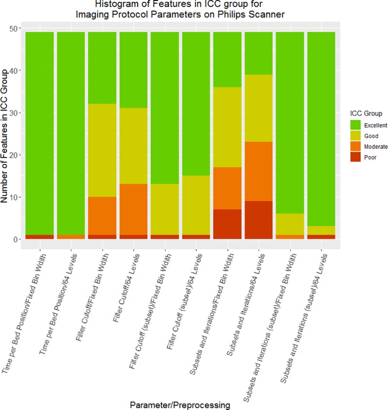
Radiomics studies require large patient cohorts, which often include patients imaged using different imaging protocols. We aimed to determine the impact of variability in imaging protocol parameters and interscanner variability using a phantom that produced feature values similar to those of patients. Positron emission tomography (PET) scans of a Hoffman brain phantom were acquired on GE Discovery 710, Siemens mCT, and Philips Vereos scanners. A standard-protocol scan was acquired on each machine, and then each parameter that could be changed was altered individually. The phantom was contoured with 10 regions of interest (ROIs). Values for 45 features with 2 different preprocessing techniques were extracted for each image. To determine the impact of each parameter on the reliability of each radiomics feature, the intraclass correlation coefficient (ICC) was calculated with the ROIs as the subjects and the parameter values as the raters. For interscanner comparisons, we compared the standard deviation of each radiomics feature value from the standard-protocol images to the standard deviation of the same radiomics feature from PET scans of 224 patients with non-small cell lung cancer. When the pixel size was resampled prior to feature extraction, all features had good reliability (ICC > 0.75) for the field of view and matrix size. The time per bed position had excellent reliability (ICC > 0.9) on all features. When the filter cutoff was restricted to values below 6 mm, all features had good reliability. Similarly, when subsets and iterations were restricted to reasonable values used in clinics, almost all features had good reliability. The average ratio of the standard deviation of features on the phantom scans to that of the NSCLC patient scans was 0.73 using fixed-bin-width preprocessing and 0.92 using 64-level preprocessing. Most radiomics feature values had at least good reliability when imaging protocol parameters were within clinically used ranges. However, interscanner variability was about equal to interpatient variability; therefore, caution must be used when combining patients scanned on equipment from different vendors in radiomics data sets.
放射组学研究需要大量的患者队列,这些队列通常包括使用不同成像方案进行成像的患者。我们旨在通过生成与患者特征值相似的特征值的体模来确定成像方案参数和扫描仪间变异性的影响。在 GE Discovery 710、Siemens mCT 和 Philips Vereos 扫描仪上对 Hoffman 脑体模进行正电子发射断层扫描(PET)扫描。在每台机器上采集标准方案扫描,然后单独改变可以更改的每个参数。使用 10 个感兴趣区域(ROI)对体模进行轮廓绘制。为每个图像提取了 2 种不同预处理技术的 45 个特征值。为了确定每个参数对每个放射组学特征的可靠性的影响,使用 ROI 作为主体,参数值作为评分者,计算了组内相关系数(ICC)。对于扫描仪间的比较,我们将标准方案图像的每个放射组学特征值的标准差与 224 例非小细胞肺癌患者的 PET 扫描的相同放射组学特征的标准差进行了比较。在提取特征之前对像素大小进行重采样时,对于视场和矩阵大小,所有特征的可靠性(ICC>0.75)都很好。对于所有特征,每个床位位置的时间都具有极好的可靠性(ICC>0.9)。当将截止值限制在 6mm 以下时,所有特征都具有良好的可靠性。类似地,当将子集和迭代限制为在临床上使用的合理值时,几乎所有特征都具有良好的可靠性。使用固定-bin-width 预处理时,体模扫描的特征标准差与非小细胞肺癌患者扫描的特征标准差的平均值比为 0.73,使用 64 级预处理时为 0.92。当成像方案参数在临床使用范围内时,大多数放射组学特征值至少具有良好的可靠性。但是,扫描仪间的变异性与患者间的变异性大致相同;因此,在放射组学数据集中组合来自不同供应商的设备扫描的患者时必须谨慎。