Department of Radiation Physics, The University of Texas MD Anderson Cancer Center, 1515 Holcombe Blvd, Houston, TX, 77030, USA.
The University of Texas Graduate School of Biomedical Sciences at Houston, 6767 Bertner Ave, Houston, TX, 77030, USA.
Sci Rep. 2017 Apr 3;7(1):588. doi: 10.1038/s41598-017-00665-z.
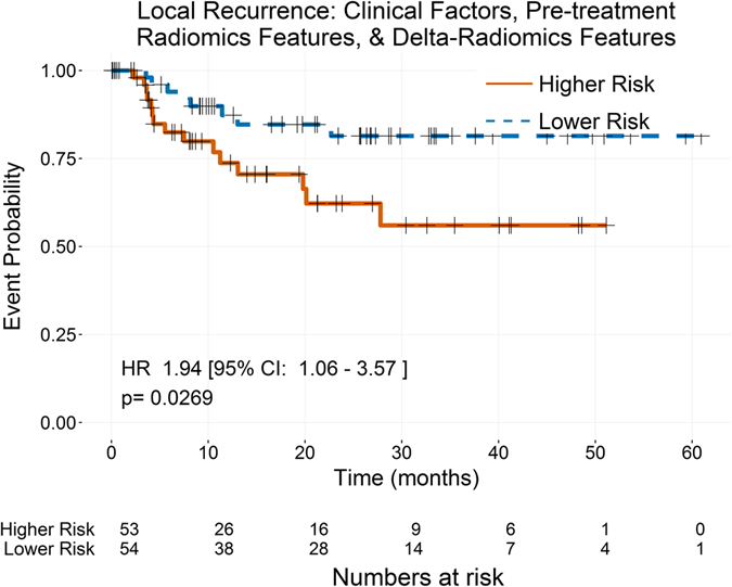
Radiomics is the use of quantitative imaging features extracted from medical images to characterize tumor pathology or heterogeneity. Features measured at pretreatment have successfully predicted patient outcomes in numerous cancer sites. This project was designed to determine whether radiomics features measured from non-small cell lung cancer (NSCLC) change during therapy and whether those features (delta-radiomics features) can improve prognostic models. Features were calculated from pretreatment and weekly intra-treatment computed tomography images for 107 patients with stage III NSCLC. Pretreatment images were used to determine feature-specific image preprocessing. Linear mixed-effects models were used to identify features that changed significantly with dose-fraction. Multivariate models were built for overall survival, distant metastases, and local recurrence using only clinical factors, clinical factors and pretreatment radiomics features, and clinical factors, pretreatment radiomics features, and delta-radiomics features. All of the radiomics features changed significantly during radiation therapy. For overall survival and distant metastases, pretreatment compactness improved the c-index. For local recurrence, pretreatment imaging features were not prognostic, while texture-strength measured at the end of treatment significantly stratified high- and low-risk patients. These results suggest radiomics features change due to radiation therapy and their values at the end of treatment may be indicators of tumor response.
放射组学是指利用从医学图像中提取的定量成像特征来描述肿瘤的病理或异质性。在许多癌症部位,在治疗前测量的特征成功地预测了患者的预后。本项目旨在确定非小细胞肺癌(NSCLC)在治疗过程中是否会发生放射组学特征的变化,以及这些特征(放射组学变化特征)是否可以改进预后模型。该研究对 107 名 III 期 NSCLC 患者的治疗前和每周的治疗中 CT 图像进行了特征测量。使用治疗前的图像来确定特定于特征的图像预处理。使用线性混合效应模型来识别与剂量-分数显著变化的特征。仅使用临床因素、临床因素和治疗前放射组学特征以及临床因素、治疗前放射组学特征和放射组学变化特征构建了用于总生存、远处转移和局部复发的多变量模型。所有的放射组学特征在放射治疗过程中都发生了显著变化。对于总生存和远处转移,治疗前的紧凑性提高了 C 指数。对于局部复发,治疗前的成像特征没有预后意义,而治疗结束时测量的纹理强度特征则显著分层了高危和低危患者。这些结果表明,放射组学特征会因放射治疗而发生变化,并且其在治疗结束时的值可能是肿瘤反应的指标。