Department of Bioengineering, School of Engineering & Applied Science, University of Pennsylvania, Philadelphia, PA 19104, USA; Center for Neuroengineering and Therapeutics, University of Pennsylvania, Philadelphia, PA 19104, USA.
Department of Bioengineering, School of Engineering & Applied Science, University of Pennsylvania, Philadelphia, PA 19104, USA; Center for Neuroengineering and Therapeutics, University of Pennsylvania, Philadelphia, PA 19104, USA.
Neuroimage Clin. 2019;23:101908. doi: 10.1016/j.nicl.2019.101908. Epub 2019 Jun 19.
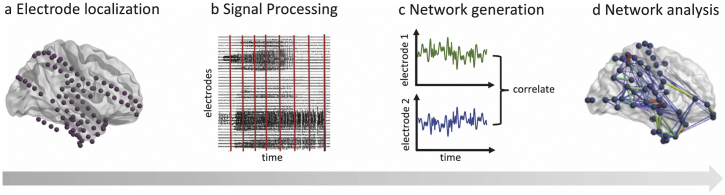
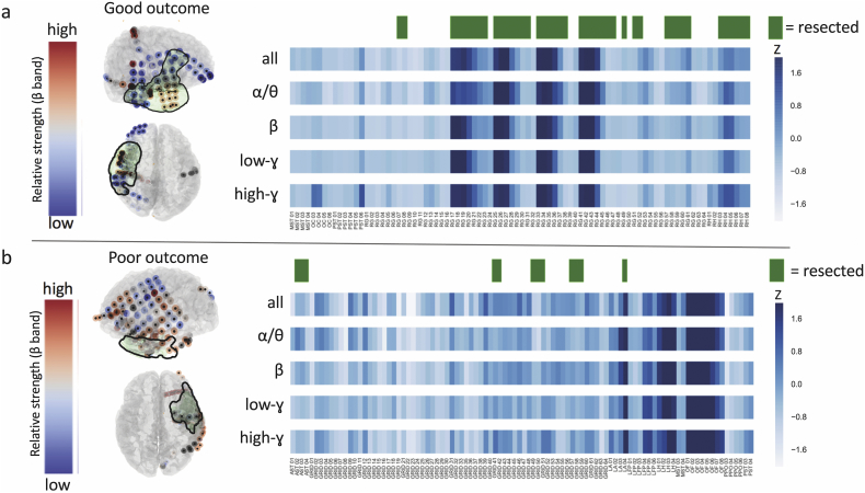
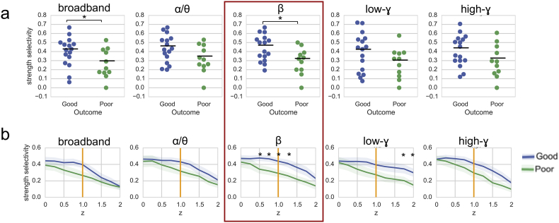
Patients with drug-resistant focal epilepsy are often candidates for invasive surgical therapies. In these patients, it is necessary to accurately localize seizure generators to ensure seizure freedom following intervention. While intracranial electroencephalography (iEEG) is the gold standard for mapping networks for surgery, this approach requires inducing and recording seizures, which may cause patient morbidity. The goal of this study is to evaluate the utility of mapping interictal (non-seizure) iEEG networks to identify targets for surgical treatment. We analyze interictal iEEG recordings and neuroimaging from 27 focal epilepsy patients treated via surgical resection. We generate interictal functional networks by calculating pairwise correlation of iEEG signals across different frequency bands. Using image coregistration and segmentation, we identify electrodes falling within surgically resected tissue (i.e. the resection zone), and compute node-level and edge-level synchrony in relation to the resection zone. We further associate these metrics with post-surgical outcomes. Greater overlap between resected electrodes and highly synchronous electrodes is associated with favorable post-surgical outcomes. Additionally, good-outcome patients have significantly higher connectivity localized within the resection zone compared to those with poorer postoperative seizure control. This finding persists following normalization by a spatially-constrained null model. This study suggests that spatially-informed interictal network synchrony measures can distinguish between good and poor post-surgical outcomes. By capturing clinically-relevant information during interictal periods, our method may ultimately reduce the need for prolonged invasive implants and provide insights into the pathophysiology of an epileptic brain. We discuss next steps for translating these findings into a prospectively useful clinical tool.
耐药性局灶性癫痫患者通常是侵袭性手术治疗的候选者。在这些患者中,有必要准确地定位癫痫发作源,以确保干预后癫痫发作得到控制。虽然颅内脑电图(iEEG)是手术中进行网络映射的金标准,但这种方法需要诱发和记录癫痫发作,这可能会导致患者发病。本研究的目的是评估利用间期(非癫痫发作)iEEG 网络进行映射以确定手术治疗靶点的效用。我们分析了 27 例经手术切除治疗的局灶性癫痫患者的间期 iEEG 记录和神经影像学数据。我们通过计算不同频段的 iEEG 信号之间的成对相关性来生成间期功能网络。通过图像配准和分割,我们识别出位于手术切除组织(即切除区)内的电极,并计算与切除区相关的节点级和边缘级同步性。我们进一步将这些指标与术后结果相关联。切除电极和高度同步电极之间的重叠越多,与术后结果越好相关。此外,与术后癫痫控制较差的患者相比,术后结果良好的患者在切除区内的局部连接性显著更高。在通过空间受限的零模型归一化后,这一发现仍然存在。本研究表明,基于空间的间期网络同步性测量可以区分术后结果良好和较差的患者。通过在间期捕获与临床相关的信息,我们的方法可能最终减少对长时间侵袭性植入物的需求,并深入了解癫痫大脑的病理生理学。我们讨论了将这些发现转化为有前景的临床工具的下一步措施。