Abid Wafaa K, Al Mukhtar Yusra H
Department of Oral and Maxillofacial Surgery, College of Dentistry, University of Mosul, Duhok, Iraq.
Dental Department of AL Khansa Hospital, Mosul, Iraq.
J Taibah Univ Med Sci. 2019 Jan 3;14(1):14-24. doi: 10.1016/j.jtumed.2018.12.001. eCollection 2019 Feb.
This study aimed to assess the repair of bone defects with or without grafts using biphasic synthetic micro-granular hyaluronic acid (HA) with β-TCP and bovine type I collagen (Osteon II Collagen) with or without either hyaluronic acid or a collagen membrane in surgically created bone defects in the tibia of rabbits.
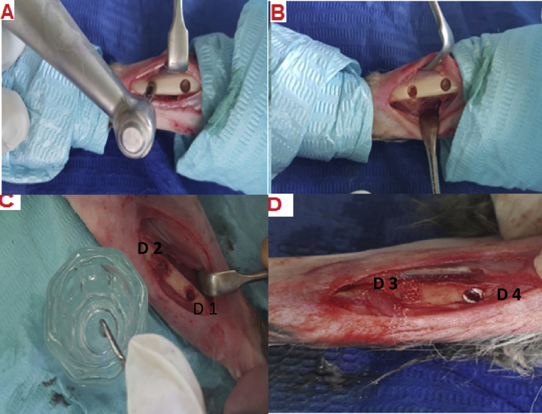
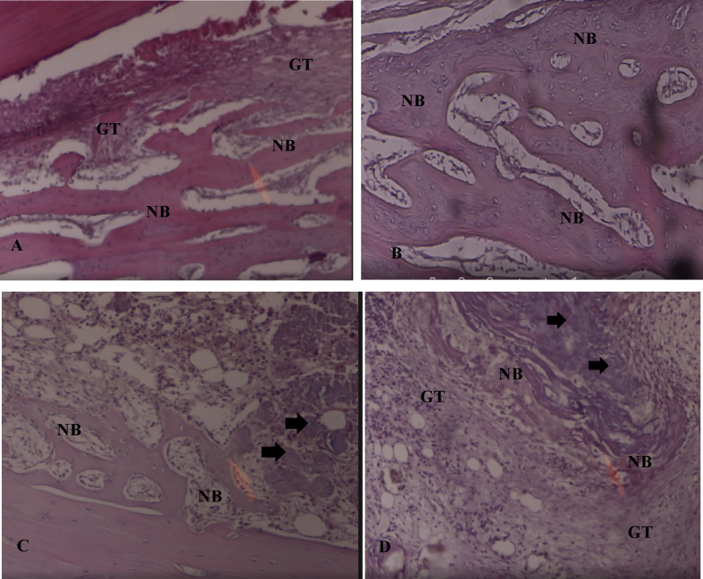
Fifteen male rabbits were divided into 3 groups, each with 5 rabbits. Two bone defects were made in each tibia. In the right tibia, the defects were either filled with clot as a control or grafted with Osteon II Collagen and hyaluronic acid. In the left tibia, the other two defects were filled with Osteon II Collagen alone or with Osteon II Collagen and a collagen membrane. The specimens were observed one, two, and four weeks after surgery. Histological examinations were used to evaluate the degree of healing according to the amount of newly formed bone.
The combination of the bone grafting biomaterial with hyaluronic acid was found to develop into the most advanced stages of the bone repair process at the second and fourth week only ( ≤ 0.05), compared to the biomaterial with a collagen membrane, as well as the other groups. On the other hand, the biomaterial in combination with a collagen membrane showed significantly more bone formation than the biomaterial alone or the control group by the fourth week.
The local application of hyaluronic acid and collagen membranes made a greater contribution to the bone repair process in the tibia of rabbits than the bone graft substitute (Osteon II Collagen) alone.
本研究旨在评估在兔胫骨手术制造的骨缺损中,使用双相合成微颗粒透明质酸(HA)与β-磷酸三钙以及牛I型胶原蛋白(骨胶原II),添加或不添加透明质酸或胶原膜的情况下,有或无移植骨时骨缺损的修复情况。
15只雄性兔子被分为3组,每组5只。每只兔子的双侧胫骨均制造两个骨缺损。右侧胫骨的缺损,一个用血凝块填充作为对照,另一个用骨胶原II和透明质酸移植。左侧胫骨的另外两个缺损,一个仅用骨胶原II填充,另一个用骨胶原II和胶原膜填充。术后1周、2周和4周观察标本。通过组织学检查,根据新形成骨的量评估愈合程度。
与含胶原膜的生物材料以及其他组相比,仅在第2周和第4周时,发现骨移植生物材料与透明质酸的组合发展到骨修复过程的最 advanced 阶段(≤0.05)。另一方面,到第4周时,与单独使用生物材料或对照组相比,与胶原膜组合的生物材料显示出明显更多的骨形成。
与单独的骨移植替代物(骨胶原II)相比,透明质酸和胶原膜的局部应用对兔胫骨的骨修复过程贡献更大。