Kubota Yoshiki, Okamoto Masahiko, Li Yang, Shiba Shintaro, Okazaki Shohei, Komatsu Shuichiro, Sakai Makoto, Kubo Nobuteru, Ohno Tatsuya, Nakano Takashi
Gunma University Heavy Ion Medical Center, Maebashi 371, Japan.
Graduate School of Medicine, Gunma University, Maebashi 371, Japan.
Cancers (Basel). 2019 Sep 27;11(10):1447. doi: 10.3390/cancers11101447.
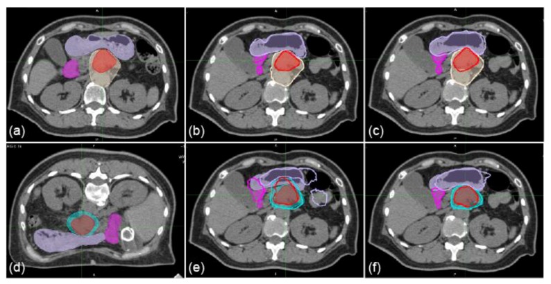
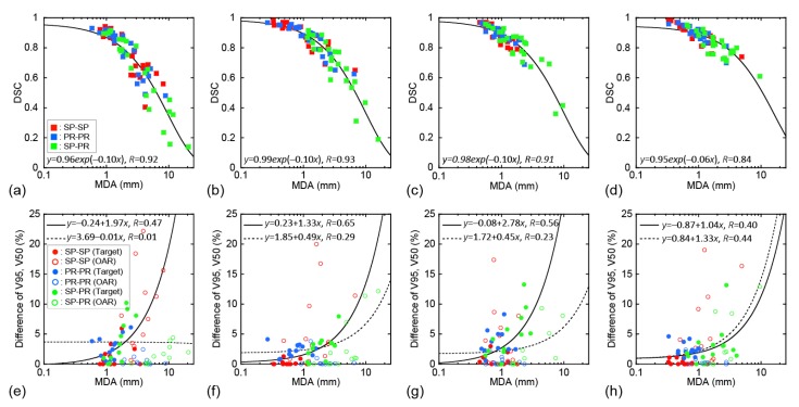
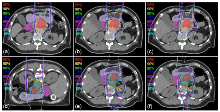
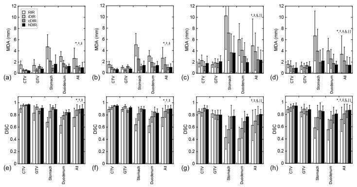
We aimed to clarify the accuracy of rigid image registration and deformable image registration (DIR) in carbon-ion radiotherapy (CIRT) for pancreatic cancer. Six patients with pancreatic cancer who were treated with passive irradiation CIRT were enrolled. Three registration patterns were evaluated: treatment planning computed tomography images (TPCT) to CT images acquired in the treatment room (IRCT) in the supine position, TPCT to IRCT in the prone position, and TPCT in the supine position to the prone position. After warping the contours of the original CT images to the destination CT images using deformation matrices from the registration, the warped delineated contours on the destination CT images were compared with the original ones using mean displacement to agreement (MDA). Four contours (clinical target volume (CTV), gross tumor volume (GTV), stomach, duodenum) and four registration algorithms (rigid image registration [RIR], intensity-based DIR [iDIR], contour-based DIR [cDIR], and a hybrid iDIR-cDIR ([hDIR]) were evaluated. The means ± standard deviation of the MDAs of all contours for RIR, iDIR, cDIR, and hDIR were 3.40 ± 3.30, 2.2 1± 2.48, 1.46 ± 1.49, and 1.46 ± 1.37 mm, respectively. There were significant differences between RIR and iDIR, and between RIR/iDIR and cDIR/hDIR. For the pancreatic cancer patient images, cDIR and hDIR had better accuracy than RIR and iDIR.
我们旨在阐明刚性图像配准和可变形图像配准(DIR)在胰腺癌碳离子放疗(CIRT)中的准确性。纳入了6例接受被动照射CIRT治疗的胰腺癌患者。评估了三种配准模式:治疗计划计算机断层扫描图像(TPCT)与在治疗室仰卧位采集的CT图像(IRCT)配准、TPCT与俯卧位的IRCT配准,以及仰卧位的TPCT与俯卧位的TPCT配准。使用配准得到的变形矩阵将原始CT图像的轮廓扭曲到目标CT图像后,使用平均位移一致性(MDA)将目标CT图像上扭曲后的勾画轮廓与原始轮廓进行比较。评估了四个轮廓(临床靶体积(CTV)、大体肿瘤体积(GTV)、胃、十二指肠)和四种配准算法(刚性图像配准[RIR]、基于强度的DIR[iDIR]、基于轮廓的DIR[cDIR]和混合iDIR - cDIR[hDIR])。RIR、iDIR、cDIR和hDIR所有轮廓的MDA均值±标准差分别为3.40±3.30、2.21±2.48、1.46±1.49和1.46±1.37mm。RIR与iDIR之间,以及RIR/iDIR与cDIR/hDIR之间存在显著差异。对于胰腺癌患者图像,cDIR和hDIR的准确性优于RIR和iDIR。