Department of Cell and Molecular Pharmacology & Experimental Therapeutics, Proteomics Center, Medical University of South Carolina, Charleston, South Carolina.
Department of Pathology and Laboratory Medicine, Medical University of South Carolina, Charleston, South Carolina.
J Mass Spectrom. 2020 Apr;55(4):e4450. doi: 10.1002/jms.4450. Epub 2020 Feb 21.
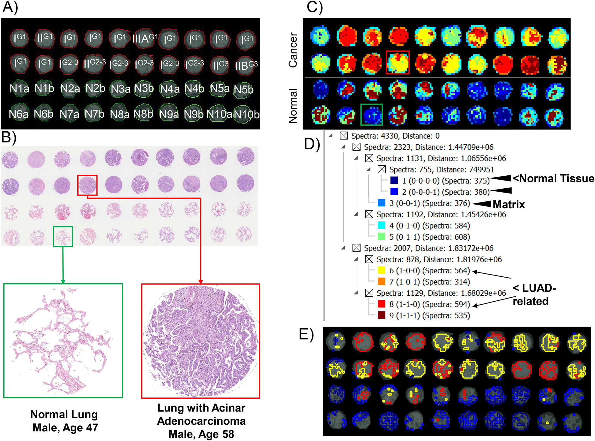
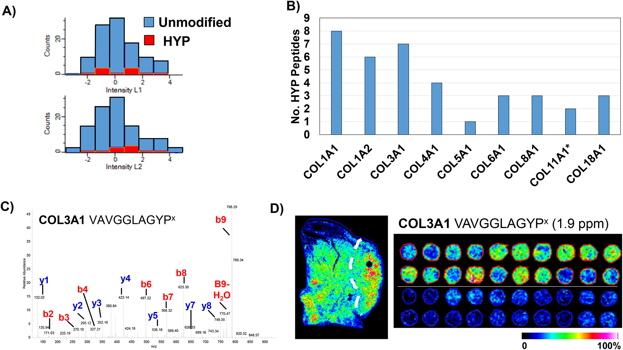
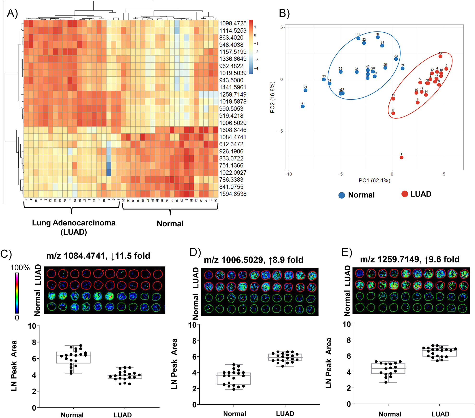
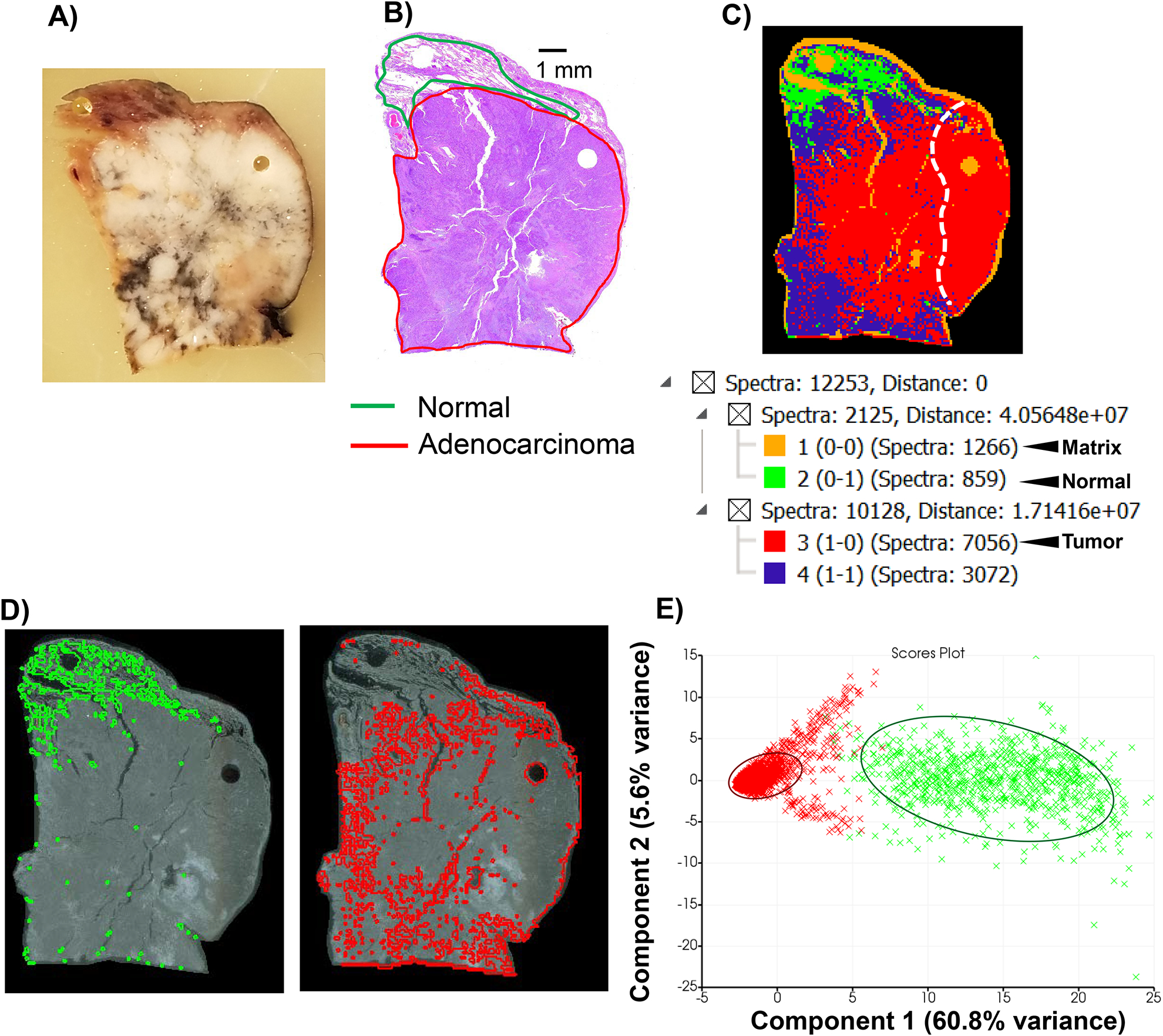
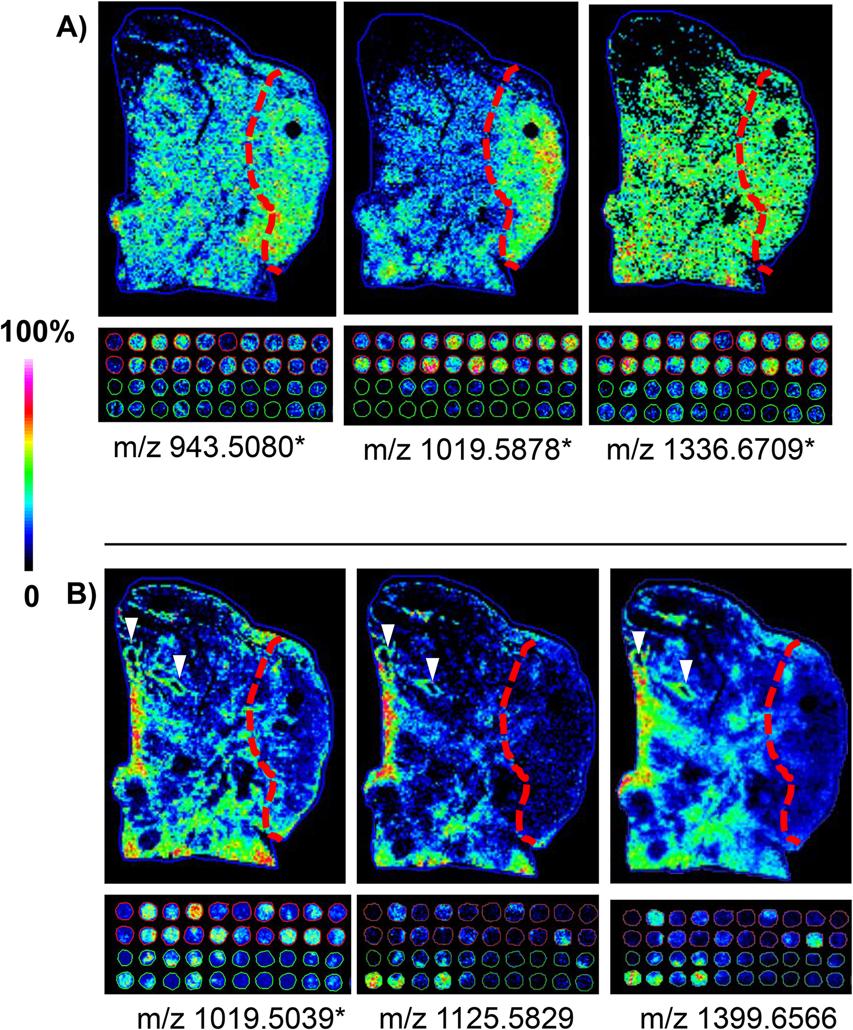
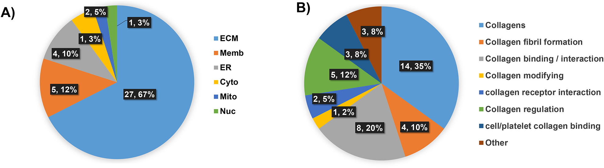
Lung adenocarcinoma (LUAD) is the second most common cancer, affecting both men and women. Fibrosis is a hallmark of LUAD occurring throughout progression with excess production of extracellular matrix (ECM) components that lead to metastatic cell processes. Understanding the ECM cues that drive LUAD progression has been limited due to a lack of tools that can access and report on ECM components within the complex tumor microenvironment. Here, we test whether low-grade LUAD can be distinguished from normal lung tissue using a novel ECM imaging mass spectrometry (ECM IMS) approach. ECM IMS analysis of a tissue microarray with 20 low-grade LUAD tissues and 20 normal lung samples from 10 patients revealed 25 peptides that could discriminate between normal and low-grade LUAD using area under the receiver-operating curve (AUC) ≥0.7, P value ≤.001. Principal component analysis demonstrated that 62.4% of the variance could be explained by sample origin from normal or low-grade tumor tissue. Additional work performed on a wedge resection with moderately differentiated LUAD demonstrated that the ECM IMS analytical approach could distinguish LUAD spectral features from spectral features of normal adjacent lung tissue. Conventional liquid chromatography with tandem mass spectrometry (LC-MS/MS) proteomics demonstrated that specific sites of hydroxylation of proline (HYP) were a main collagen post translational modification that was readily detected in LUAD. A distinct peptide from collagen 3A1 modified by HYP was increased 3.5 fold in low-grade LUAD compared with normal lung tissue (AUC 0.914, P value <.001). This suggests that regulation of collagen proline hydroxylation could be an important process during early LUAD fibrotic deposition. ECM IMS is a useful tool that may be used to define fibrotic deposition in low-grade LUAD.
肺腺癌(LUAD)是第二大常见癌症,影响男性和女性。纤维化是 LUAD 发生的标志,贯穿于整个进展过程,导致细胞外基质(ECM)成分过度产生,从而导致转移性细胞过程。由于缺乏可以访问和报告复杂肿瘤微环境中 ECM 成分的工具,因此对驱动 LUAD 进展的 ECM 线索的理解受到限制。在这里,我们测试了使用新型 ECM 成像质谱(ECM IMS)方法是否可以区分低级别 LUAD 和正常肺组织。对来自 10 名患者的 20 个低级别 LUAD 组织和 20 个正常肺样本的组织微阵列进行的 ECM IMS 分析显示,使用 AUC≥0.7、P 值≤0.001 的 25 个肽可以区分正常和低级别 LUAD。主成分分析表明,62.4%的方差可以由样本来源(正常或低级别肿瘤组织)来解释。在具有中度分化 LUAD 的楔形切除术中进行的额外工作表明,ECM IMS 分析方法可以区分 LUAD 的光谱特征与正常相邻肺组织的光谱特征。传统的液相色谱-串联质谱(LC-MS/MS)蛋白质组学表明,脯氨酸(HYP)的特定羟化部位是一种主要的胶原蛋白翻译后修饰,在 LUAD 中很容易检测到。与正常肺组织相比,来自胶原 3A1 的经 HYP 修饰的独特肽在低级别 LUAD 中增加了 3.5 倍(AUC 0.914,P 值<.001)。这表明胶原脯氨酸羟化的调节可能是 LUAD 早期纤维化沉积过程中的一个重要过程。ECM IMS 是一种有用的工具,可用于定义低级别 LUAD 中的纤维化沉积。