Kalinovic Sanela, Oelze Matthias, Kröller-Schön Swenja, Steven Sebastian, Vujacic-Mirski Ksenija, Kvandová Miroslava, Schmal Isabella, Al Zuabi Ahmad, Münzel Thomas, Daiber Andreas
Center for Cardiology, Department of Cardiology, Molecular Cardiology, University Medical Center, 55131 Mainz, Germany.
Partner Site Rhine-Main, German Center for Cardiovascular Research (DZHK), Langenbeckstr. 1, 55131 Mainz, Germany.
Antioxidants (Basel). 2019 Oct 28;8(11):514. doi: 10.3390/antiox8110514.
Reactive oxygen and nitrogen species (RONS such as HO, nitric oxide) are generated within the organism. Whereas physiological formation rates confer redox regulation of essential cellular functions and provide the basis for adaptive stress responses, their excessive formation contributes to impaired cellular function or even cell death, organ dysfunction and severe disease phenotypes of the entire organism. Therefore, quantification of RONS formation and knowledge of their tissue/cell/compartment-specific distribution is of great biological and clinical importance.
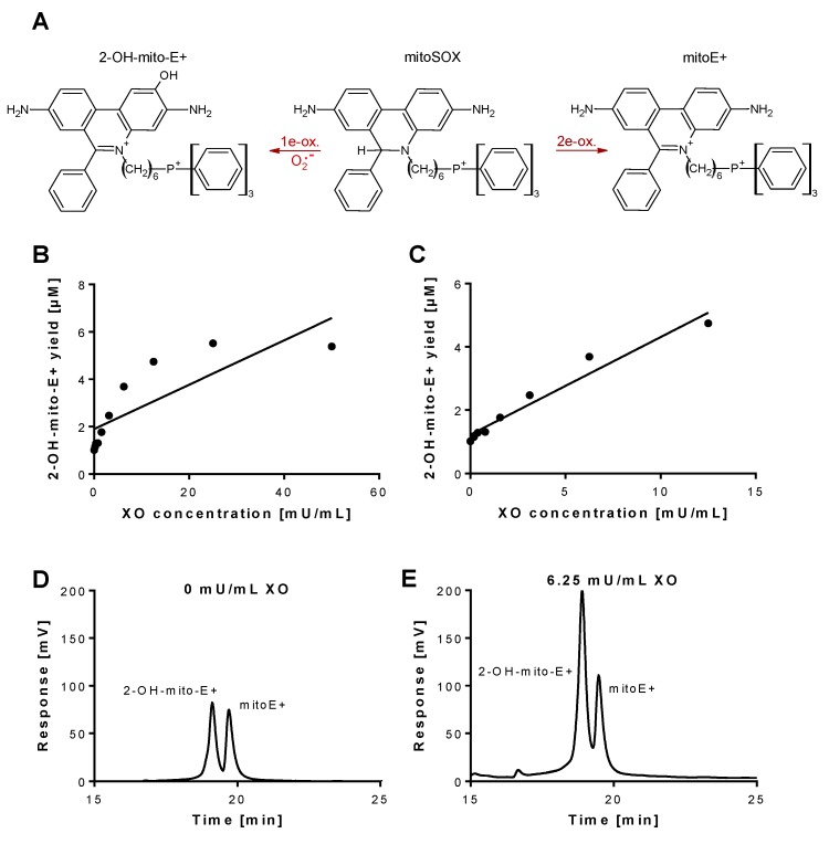
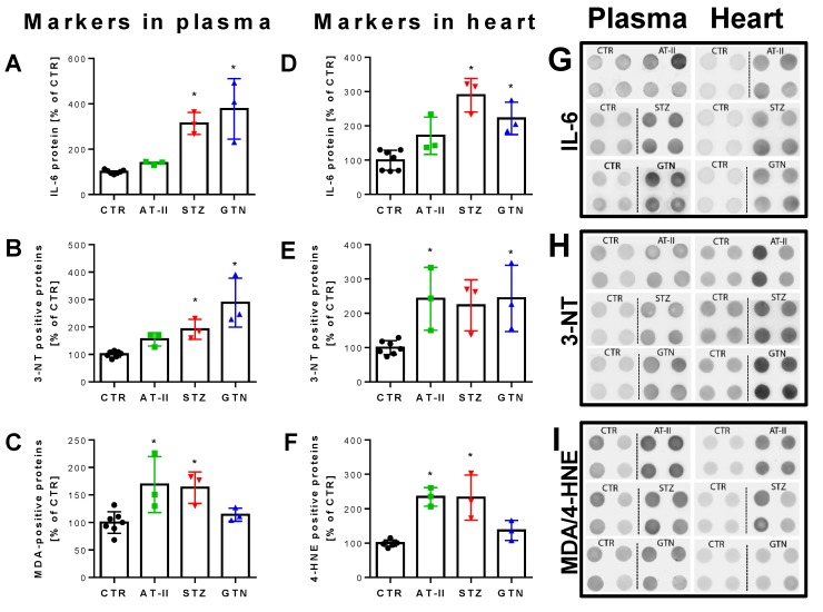
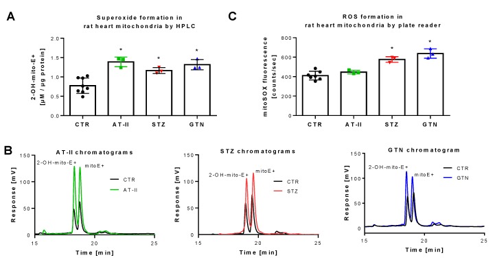
Here, we used a high-performance/pressure liquid chromatography (HPLC) assay to quantify the superoxide-specific oxidation product of the mitochondria-targeted fluorescence dye triphenylphosphonium-linked hydroethidium (mitoSOX) in biochemical systems and three animal models with established oxidative stress. Type 1 diabetes (single injection of streptozotocin), hypertension (infusion of angiotensin-II for 7 days) and nitrate tolerance (infusion of nitroglycerin for 4 days) was induced in male Wistar rats.
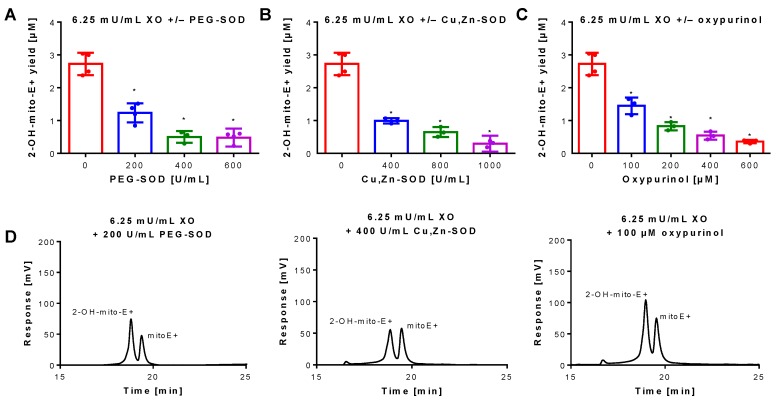
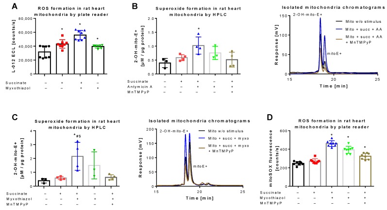
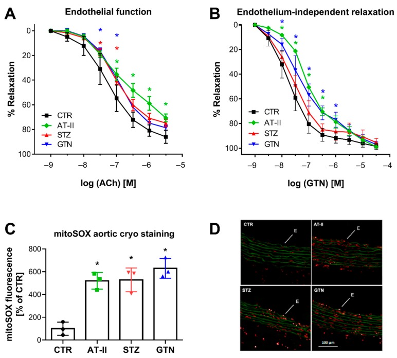
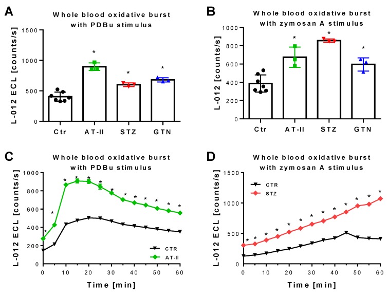
The usefulness of mitoSOX/HPLC for quantification of mitochondrial superoxide was confirmed by xanthine oxidase activity as well as isolated stimulated rat heart mitochondria in the presence or absence of superoxide scavengers. Vascular function was assessed by isometric tension methodology and was impaired in the rat models of oxidative stress. Vascular dysfunction correlated with increased mitoSOX oxidation but also classical RONS detection assays as well as typical markers of oxidative stress.
mitoSOX/HPLC represents a valid method for detection of mitochondrial superoxide formation in tissues of different animal disease models and correlates well with functional parameters and other markers of oxidative stress.
活性氧和氮物种(如羟基自由基、一氧化氮等活性氧氮化物)在生物体内产生。生理生成速率可对基本细胞功能进行氧化还原调节,并为适应性应激反应提供基础,而其过度生成则会导致细胞功能受损甚至细胞死亡、器官功能障碍以及整个生物体的严重疾病表型。因此,活性氧氮化物生成的定量分析及其在组织/细胞/区室特异性分布的了解具有重要的生物学和临床意义。
在此,我们使用高效/高压液相色谱(HPLC)分析法,对线粒体靶向荧光染料三苯基鏻连接的氢乙锭(mitoSOX)在生化系统和三种已建立氧化应激的动物模型中的超氧化物特异性氧化产物进行定量。在雄性Wistar大鼠中诱导1型糖尿病(单次注射链脲佐菌素)、高血压(输注血管紧张素-II 7天)和硝酸盐耐受性(输注硝酸甘油4天)。
黄嘌呤氧化酶活性以及在有或没有超氧化物清除剂存在的情况下分离的受刺激大鼠心脏线粒体证实了mitoSOX/HPLC用于定量线粒体超氧化物的有效性。通过等长张力法评估血管功能,在氧化应激大鼠模型中血管功能受损。血管功能障碍与mitoSOX氧化增加相关,但也与经典的活性氧氮化物检测方法以及氧化应激的典型标志物相关。
mitoSOX/HPLC是检测不同动物疾病模型组织中线粒体超氧化物生成的有效方法,并且与功能参数和氧化应激的其他标志物相关性良好。