Sato Wataru, Kochiyama Takanori, Uono Shota, Yoshimura Sayaka, Kubota Yasutaka, Sawada Reiko, Sakihama Morimitsu, Toichi Motomi
Kokoro Research Center, Kyoto University, Kyoto, Japan.
Brain Activity Imaging Center, ATR-Promotions, Inc., Kyoto, Japan.
Front Hum Neurosci. 2019 Oct 18;13:351. doi: 10.3389/fnhum.2019.00351. eCollection 2019.
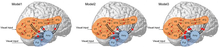
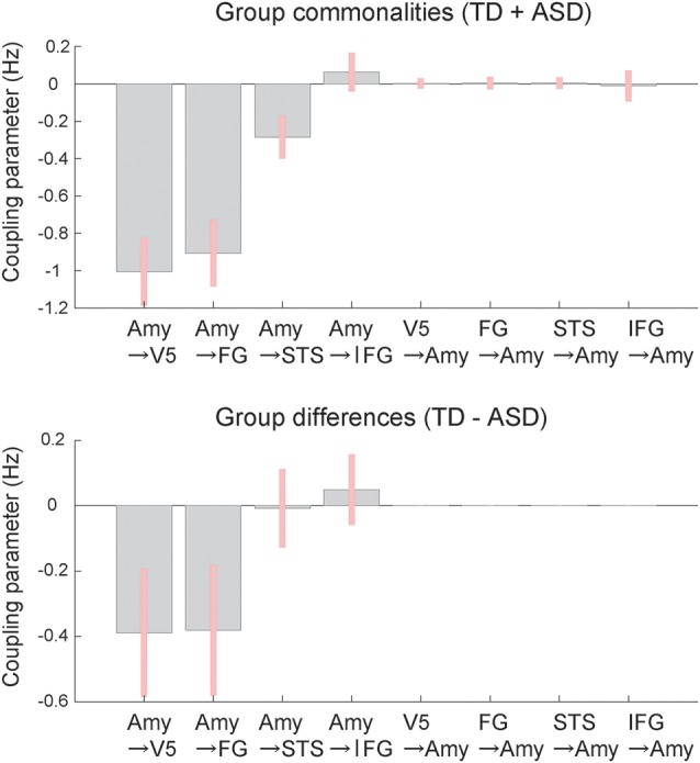
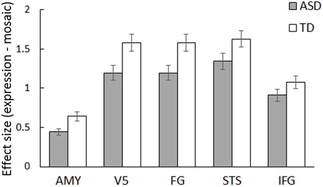
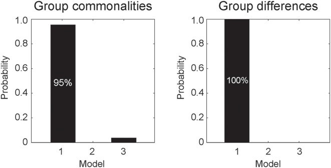
Atypical reciprocal social interactions involving emotional facial expressions are a core clinical feature of autism spectrum disorder (ASD). Previous functional magnetic resonance imaging (fMRI) studies have demonstrated that some social brain regions, including subcortical (e.g., amygdala) and neocortical regions (e.g., fusiform gyrus, FG) are less activated during the processing of facial expression stimuli in individuals with ASD. However, the functional networking patterns between the subcortical and cortical regions in processing emotional facial expressions remain unclear. We investigated this issue in ASD ( = 31) and typically developing (TD; = 31) individuals using fMRI. Participants viewed dynamic facial expressions of anger and happiness and their corresponding mosaic images. Regional brain activity analysis revealed reduced activation of several social brain regions, including the amygdala, in the ASD group compared with the TD group in response to dynamic facial expressions vs. dynamic mosaics ( < 0.05, = 0.19). Dynamic causal modeling (DCM) analyses were then used to compare models with forward, backward, and bi-directional effective connectivity between the amygdala and neocortical networks. The results revealed that: (1) the model with effective connectivity from the amygdala to the neocortex best fit the data of both groups; and (2) the same model best accounted for group differences. Coupling parameter (i.e., effective connectivity) analyses showed that the modulatory effects of dynamic facial processing were substantially weaker in the ASD group than in the TD group. These findings suggest that atypical modulation from the amygdala to the neocortex underlies impairment in social interaction involving dynamic facial expressions in individuals with ASD.
涉及情感面部表情的非典型互惠社交互动是自闭症谱系障碍(ASD)的核心临床特征。先前的功能磁共振成像(fMRI)研究表明,在处理面部表情刺激时,一些社会脑区,包括皮层下区域(如杏仁核)和新皮层区域(如梭状回,FG),在患有ASD的个体中激活程度较低。然而,在处理情感面部表情时,皮层下和皮层区域之间的功能网络模式仍不清楚。我们使用fMRI在ASD患者(n = 31)和正常发育(TD;n = 31)个体中研究了这个问题。参与者观看了愤怒和快乐的动态面部表情及其相应的镶嵌图像。区域脑活动分析显示,与TD组相比,ASD组在对动态面部表情与动态镶嵌图像的反应中,包括杏仁核在内的几个社会脑区的激活减少(p < 0.05,η² = 0.19)。然后使用动态因果模型(DCM)分析来比较杏仁核和新皮层网络之间具有正向、反向和双向有效连接的模型。结果显示:(1)从杏仁核到新皮层具有有效连接的模型最适合两组的数据;(2)相同的模型最能解释组间差异。耦合参数(即有效连接)分析表明,ASD组中动态面部处理的调节作用明显弱于TD组。这些发现表明,杏仁核到新皮层的非典型调节是导致ASD个体在涉及动态面部表情的社交互动中受损的基础。