Physical Education and Sports Science Academic Group, National Institute of Education, Nanyang Technological University, Singapore, Singapore.
Centre for Research and Development in Learning, Nanyang Technological University, Singapore, Singapore.
Brain Imaging Behav. 2023 Apr;17(2):257-269. doi: 10.1007/s11682-022-00754-2. Epub 2023 Jan 12.
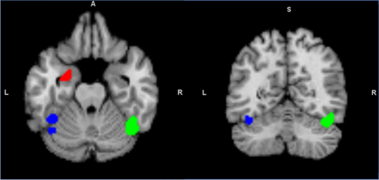
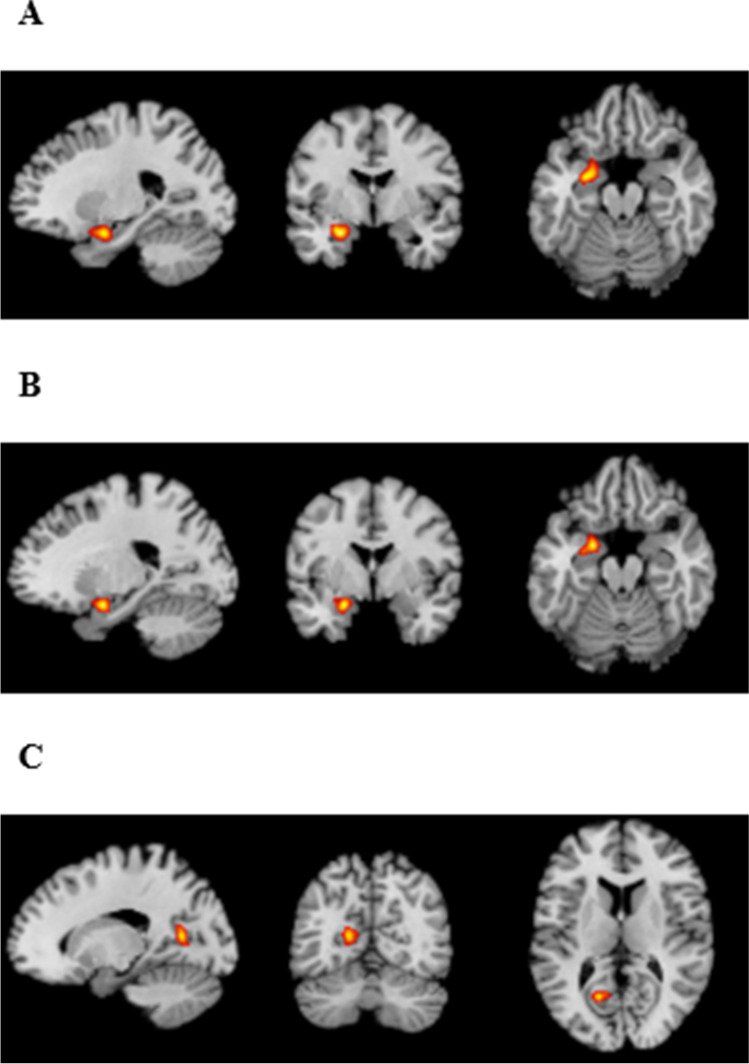
Social and non-social deficits in autism spectrum disorders (ASD) persist into adulthood and may share common regions of aberrant neural activations. The current meta-analysis investigated activation differences between ASD and neurotypical controls irrespective of task type. Activation likelihood estimation meta-analyses were performed to examine consistent hypo-activated and/or hyper-activated regions for all tasks combined, and for social and non-social tasks separately; meta-analytic connectivity modelling and behavioral/paradigm analyses were performed to examine co-activated regions and associated behaviors. One hundred studies (mean age range = 18-41 years) were included. For all tasks combined, the ASD group showed significant (p < .05) hypo-activation in one cluster around the left amygdala (peak - 26, -2, -20, volume = 1336 mm, maximum ALE = 0.0327), and this cluster co-activated with two other clusters around the right cerebellum (peak 42, -56, -22, volume = 2560mm, maximum ALE = 0.049) Lobule VI/Crus I and the left fusiform gyrus (BA47) (peak - 42, -46, -18, volume = 1616 mm, maximum ALE = 0.046) and left cerebellum (peak - 42, -58, -20, volume = 1616mm, maximum ALE = 0.033) Lobule VI/Crus I. While the left amygdala was associated with negative emotion (fear) (z = 3.047), the left fusiform gyrus/cerebellum Lobule VI/Crus I cluster was associated with language semantics (z = 3.724) and action observation (z = 3.077). These findings highlight the left amygdala as a region consistently hypo-activated in ASD and suggest the potential involvement of fusiform gyrus and cerebellum in social cognition in ASD. Future research should further elucidate if and how amygdala-fusiform/cerebellar connectivity relates to social and non-social cognition in adults with ASD.
自闭症谱系障碍(ASD)患者的社交和非社交缺陷会持续到成年期,并且可能存在共同的异常神经激活区域。本研究旨在对 ASD 患者和神经典型对照者进行任务类型无关的神经激活差异进行元分析。我们采用激活似然估计元分析来检验所有任务组合以及社交和非社交任务的一致性低激活和/或高激活区域;采用元分析连接组学和行为/范式分析来检验共同激活区域和相关行为。共纳入 100 项研究(平均年龄范围为 18-41 岁)。对于所有任务的组合,ASD 组在左侧杏仁核周围有一个显著的(p<0.05)低激活簇(峰值为-26,-2,-20,体积为 1336mm,最大 ALE 值为 0.0327),该簇与右侧小脑的两个其他簇共同激活(峰值为 42,-56,-22,体积为 2560mm,最大 ALE 值为 0.049)小脑 VI/Crus I 和左侧梭状回(BA47)(峰值为-42,-46,-18,体积为 1616mm,最大 ALE 值为 0.046)和左侧小脑(峰值为-42,-58,-20,体积为 1616mm,最大 ALE 值为 0.033)小脑 VI/Crus I。左侧杏仁核与负性情绪(恐惧)相关(z=3.047),而左侧梭状回/小脑 VI/Crus I 簇与语言语义(z=3.724)和动作观察(z=3.077)相关。这些发现强调了左侧杏仁核在 ASD 中是一致低激活的区域,并提示了梭状回和小脑在 ASD 患者社会认知中的潜在作用。未来的研究应该进一步阐明 ASD 成人的杏仁核-梭状回/小脑连接与社交和非社交认知的关系。