• 文献检索
  • 文档翻译
  • 深度研究
  • 学术资讯
  • Suppr Zotero 插件Zotero 插件
  • 邀请有礼
  • 套餐&价格
  • 历史记录
应用&插件
Suppr Zotero 插件Zotero 插件浏览器插件Mac 客户端Windows 客户端微信小程序
定价
高级版会员购买积分包购买API积分包
服务
文献检索文档翻译深度研究API 文档MCP 服务
关于我们
关于 Suppr公司介绍联系我们用户协议隐私条款
关注我们

Suppr 超能文献

核心技术专利:CN118964589B侵权必究
粤ICP备2023148730 号-1Suppr @ 2026

文献检索

告别复杂PubMed语法,用中文像聊天一样搜索,搜遍4000万医学文献。AI智能推荐,让科研检索更轻松。

立即免费搜索

文件翻译

保留排版,准确专业,支持PDF/Word/PPT等文件格式,支持 12+语言互译。

免费翻译文档

深度研究

AI帮你快速写综述,25分钟生成高质量综述,智能提取关键信息,辅助科研写作。

立即免费体验

增强和统一的常见小鼠脑图谱解剖学标记。

Enhanced and unified anatomical labeling for a common mouse brain atlas.

机构信息

Department of Neural and Behavioral Sciences, College of Medicine, Penn State University, Hershey, PA, USA.

Department of Pathology, College of Medicine, Penn State University, Hershey, PA, USA.

出版信息

Nat Commun. 2019 Nov 7;10(1):5067. doi: 10.1038/s41467-019-13057-w.

DOI:10.1038/s41467-019-13057-w
PMID:31699990
原文链接:https://pmc.ncbi.nlm.nih.gov/articles/PMC6838086/
Abstract

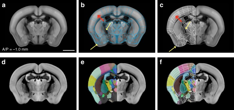
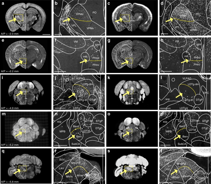
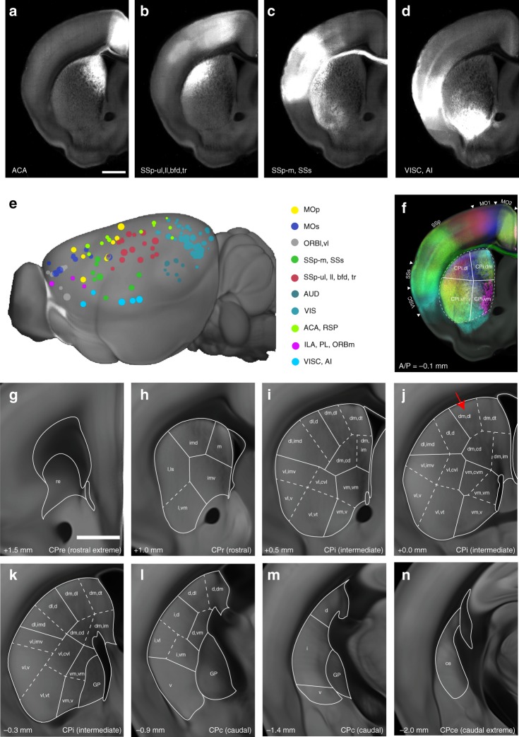
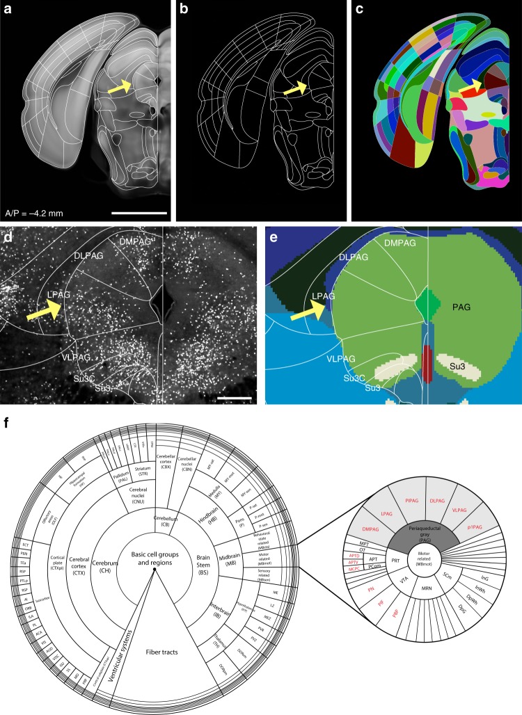
Anatomical atlases in standard coordinates are necessary for the interpretation and integration of research findings in a common spatial context. However, the two most-used mouse brain atlases, the Franklin-Paxinos (FP) and the common coordinate framework (CCF) from the Allen Institute for Brain Science, have accumulated inconsistencies in anatomical delineations and nomenclature, creating confusion among neuroscientists. To overcome these issues, we adopt here the FP labels into the CCF to merge the labels in the single atlas framework. We use cell type-specific transgenic mice and an MRI atlas to adjust and further segment our labels. Moreover, detailed segmentations are added to the dorsal striatum using cortico-striatal connectivity data. Lastly, we digitize our anatomical labels based on the Allen ontology, create a web-interface for visualization, and provide tools for comprehensive comparisons between the CCF and FP labels. Our open-source labels signify a key step towards a unified mouse brain atlas.

摘要

标准坐标的解剖图谱对于在共同的空间背景下解释和整合研究结果是必要的。然而,两个最常用的小鼠脑图谱,即富兰克林-帕克斯诺斯(FP)图谱和艾伦脑科学研究所的共同坐标框架(CCF),在解剖划分和命名上积累了不一致之处,给神经科学家带来了困惑。为了解决这些问题,我们在这里采用 FP 标签到 CCF 中,将标签合并到单个图谱框架中。我们使用细胞类型特异性转基因小鼠和 MRI 图谱来调整和进一步分割我们的标签。此外,我们使用皮质-纹状体连接数据为背侧纹状体添加了详细的分割。最后,我们基于艾伦本体对我们的解剖标签进行数字化,创建了一个可视化的网络界面,并提供了用于 CCF 和 FP 标签之间全面比较的工具。我们的开源标签标志着迈向统一的小鼠脑图谱迈出了关键的一步。

https://cdn.ncbi.nlm.nih.gov/pmc/blobs/d9c9/6838086/0c9416245482/41467_2019_13057_Fig6_HTML.jpg
https://cdn.ncbi.nlm.nih.gov/pmc/blobs/d9c9/6838086/135857820949/41467_2019_13057_Fig1_HTML.jpg
https://cdn.ncbi.nlm.nih.gov/pmc/blobs/d9c9/6838086/b91ac7373f58/41467_2019_13057_Fig2_HTML.jpg
https://cdn.ncbi.nlm.nih.gov/pmc/blobs/d9c9/6838086/2b4d465c552c/41467_2019_13057_Fig3_HTML.jpg
https://cdn.ncbi.nlm.nih.gov/pmc/blobs/d9c9/6838086/b19300d9b25c/41467_2019_13057_Fig4_HTML.jpg
https://cdn.ncbi.nlm.nih.gov/pmc/blobs/d9c9/6838086/e1b1f519aca6/41467_2019_13057_Fig5_HTML.jpg
https://cdn.ncbi.nlm.nih.gov/pmc/blobs/d9c9/6838086/0c9416245482/41467_2019_13057_Fig6_HTML.jpg

相似文献

1
Enhanced and unified anatomical labeling for a common mouse brain atlas.增强和统一的常见小鼠脑图谱解剖学标记。
Nat Commun. 2019 Nov 7;10(1):5067. doi: 10.1038/s41467-019-13057-w.
2
A stereotaxic MRI template set of mouse brain with fine sub-anatomical delineations: Application to MEMRI studies of 5XFAD mice.一套具有精细亚解剖结构描绘的小鼠脑立体定向MRI模板集:应用于5XFAD小鼠的磁共振弹性成像研究。
Magn Reson Imaging. 2019 Apr;57:83-94. doi: 10.1016/j.mri.2018.10.014. Epub 2018 Oct 22.
3
Connecting imaging mass spectrometry and magnetic resonance imaging-based anatomical atlases for automated anatomical interpretation and differential analysis.将影像质谱和基于磁共振成像的解剖图谱连接起来,用于自动解剖解释和差异分析。
Biochim Biophys Acta Proteins Proteom. 2017 Jul;1865(7):967-977. doi: 10.1016/j.bbapap.2017.02.016. Epub 2017 Feb 27.
4
AtOM, an ontology model to standardize use of brain atlases in tools, workflows, and data infrastructures.AtOM,一种本体模型,用于在工具、工作流程和数据基础架构中标准化脑图谱的使用。
Sci Data. 2023 Jul 26;10(1):486. doi: 10.1038/s41597-023-02389-4.
5
Constructing and optimizing 3D atlases from 2D data with application to the developing mouse brain.从二维数据构建和优化 3D 图谱及其在发育中老鼠大脑中的应用。
Elife. 2021 Feb 11;10:e61408. doi: 10.7554/eLife.61408.
6
A rat brain MRI template with digital stereotaxic atlas of fine anatomical delineations in paxinos space and its automated application in voxel-wise analysis.帕克斯诺空间精细解剖标记大鼠脑 MRI 模板及其在体素分析中的自动应用
Hum Brain Mapp. 2013 Jun;34(6):1306-18. doi: 10.1002/hbm.21511. Epub 2012 Jan 30.
7
Neuroinformatics of the Allen Mouse Brain Connectivity Atlas.艾伦小鼠脑连接图谱的神经信息学
Methods. 2015 Feb;73:4-17. doi: 10.1016/j.ymeth.2014.12.013. Epub 2014 Dec 20.
8
Common atlas format and 3D brain atlas reconstructor: infrastructure for constructing 3D brain atlases.通用图谱格式和 3D 脑图谱重构器:构建 3D 脑图谱的基础架构。
Neuroinformatics. 2012 Apr;10(2):181-97. doi: 10.1007/s12021-011-9138-6.
9
The Allen Mouse Brain Common Coordinate Framework: A 3D Reference Atlas.艾伦鼠脑通用坐标系:一个 3D 参考图谱。
Cell. 2020 May 14;181(4):936-953.e20. doi: 10.1016/j.cell.2020.04.007. Epub 2020 May 7.
10
Developmental Mouse Brain Common Coordinate Framework.发育中的小鼠大脑通用坐标框架
bioRxiv. 2023 Sep 15:2023.09.14.557789. doi: 10.1101/2023.09.14.557789.

引用本文的文献

1
An extended and improved CCFv3 annotation and Nissl atlas of the entire mouse brain.扩展并改进的整个小鼠脑CCFv3注释和尼氏图谱。
Imaging Neurosci (Camb). 2025 May 21;3. doi: 10.1162/imag_a_00565. eCollection 2025.
2
Differential impacts of social isolation and enriched environment on multi-sensory brain-wide functionality and network segregation.社会隔离和丰富环境对多感官全脑功能及网络分离的不同影响。
Nat Commun. 2025 Jul 31;16(1):7036. doi: 10.1038/s41467-025-62253-4.
3
A SMARTTR workflow for multi-ensemble atlas mapping and brain-wide network analysis.

本文引用的文献

1
A repeated molecular architecture across thalamic pathways.丘脑通路上的重复分子结构。
Nat Neurosci. 2019 Nov;22(11):1925-1935. doi: 10.1038/s41593-019-0483-3. Epub 2019 Sep 16.
2
Processing Pipeline for Atlas-Based Imaging Data Analysis of Structural and Functional Mouse Brain MRI (AIDAmri).基于图谱的小鼠脑结构和功能磁共振成像(AIDAmri)数据分析处理流程
Front Neuroinform. 2019 Jun 4;13:42. doi: 10.3389/fninf.2019.00042. eCollection 2019.
3
An active texture-based digital atlas enables automated mapping of structures and markers across brains.
一种用于多集合图谱映射和全脑网络分析的SMARTTR工作流程。
Elife. 2025 Jul 25;13:RP101327. doi: 10.7554/eLife.101327.
4
Primary auditory thalamus relays directly to cortical layer 1 interneurons.初级听觉丘脑直接投射至皮质第1层中间神经元。
iScience. 2025 Jun 24;28(8):112652. doi: 10.1016/j.isci.2025.112652. eCollection 2025 Aug 15.
5
A mouse brain stereotaxic topographic atlas with isotropic 1-μm resolution.具有各向同性1微米分辨率的小鼠脑立体定位地形图集。
Nature. 2025 Jul 2. doi: 10.1038/s41586-025-09211-8.
6
GPR43 regulates mitochondrial apoptosis through the cyclophilin D pathway in Alzheimer's disease.GPR43通过亲环素D途径调节阿尔茨海默病中的线粒体凋亡。
Mol Med. 2025 Jun 4;31(1):219. doi: 10.1186/s10020-025-01269-4.
7
Modeling functional connectivity with learning and memory in a mouse model of Alzheimer's disease.在阿尔茨海默病小鼠模型中建立与学习和记忆相关的功能连接模型。
Front Neuroimaging. 2025 Apr 25;4:1558759. doi: 10.3389/fnimg.2025.1558759. eCollection 2025.
8
The Duke Mouse Brain Atlas: MRI and light sheet microscopy stereotaxic atlas of the mouse brain.《杜克小鼠脑图谱:小鼠脑的磁共振成像和光片显微镜立体定位图谱》
Sci Adv. 2025 May 2;11(18):eadq8089. doi: 10.1126/sciadv.adq8089. Epub 2025 Apr 30.
9
Modular strategies for spatial mapping of diverse cell type data of the mouse brain.小鼠大脑多种细胞类型数据空间映射的模块化策略。
Res Sq. 2025 Apr 9:rs.3.rs-6289741. doi: 10.21203/rs.3.rs-6289741/v1.
10
A Comprehensive Atlas of Cell Type Density Patterns and Their Role in Brain Organization.《细胞类型密度模式综合图谱及其在脑组织结构中的作用》
bioRxiv. 2025 Mar 20:2024.10.02.615922. doi: 10.1101/2024.10.02.615922.
基于活动纹理的数字图谱可实现大脑结构和标记物的自动配准。
Nat Methods. 2019 Apr;16(4):341-350. doi: 10.1038/s41592-019-0328-8. Epub 2019 Mar 11.
4
A high-throughput neurohistological pipeline for brain-wide mesoscale connectivity mapping of the common marmoset.高通量神经组织学流水线用于常见绒猴全脑介观连接图谱绘制。
Elife. 2019 Feb 5;8:e40042. doi: 10.7554/eLife.40042.
5
Automatic navigation system for the mouse brain.鼠标脑自动导航系统。
J Comp Neurol. 2019 Sep 1;527(13):2200-2211. doi: 10.1002/cne.24635. Epub 2019 Feb 24.
6
Whole mouse brain reconstruction and registration to a reference atlas with standard histochemical processing of coronal sections.对经过标准组织化学处理的冠状切片进行全鼠脑重建和与参考图谱配准。
J Comp Neurol. 2019 Sep 1;527(13):2170-2178. doi: 10.1002/cne.24602. Epub 2019 Jan 2.
7
A Cell Atlas for the Mouse Brain.小鼠大脑细胞图谱。
Front Neuroinform. 2018 Nov 28;12:84. doi: 10.3389/fninf.2018.00084. eCollection 2018.
8
Navigating the Murine Brain: Toward Best Practices for Determining and Documenting Neuroanatomical Locations in Experimental Studies.探索小鼠大脑:实验研究中确定和记录神经解剖位置的最佳实践指南
Front Neuroanat. 2018 Nov 2;12:82. doi: 10.3389/fnana.2018.00082. eCollection 2018.
9
Integration of gene expression and brain-wide connectivity reveals the multiscale organization of mouse hippocampal networks.基因表达与全脑连接的整合揭示了小鼠海马网络的多尺度组织。
Nat Neurosci. 2018 Nov;21(11):1628-1643. doi: 10.1038/s41593-018-0241-y. Epub 2018 Oct 8.
10
Topographic precision in sensory and motor corticostriatal projections varies across cell type and cortical area.感觉运动皮质纹状体投射的地形精度因细胞类型和皮质区域而异。
Nat Commun. 2018 Sep 3;9(1):3549. doi: 10.1038/s41467-018-05780-7.