• 文献检索
  • 文档翻译
  • 深度研究
  • 学术资讯
  • Suppr Zotero 插件Zotero 插件
  • 邀请有礼
  • 套餐&价格
  • 历史记录
应用&插件
Suppr Zotero 插件Zotero 插件浏览器插件Mac 客户端Windows 客户端微信小程序
定价
高级版会员购买积分包购买API积分包
服务
文献检索文档翻译深度研究API 文档MCP 服务
关于我们
关于 Suppr公司介绍联系我们用户协议隐私条款
关注我们

Suppr 超能文献

核心技术专利:CN118964589B侵权必究
粤ICP备2023148730 号-1Suppr @ 2026

文献检索

告别复杂PubMed语法,用中文像聊天一样搜索,搜遍4000万医学文献。AI智能推荐,让科研检索更轻松。

立即免费搜索

文件翻译

保留排版,准确专业,支持PDF/Word/PPT等文件格式,支持 12+语言互译。

免费翻译文档

深度研究

AI帮你快速写综述,25分钟生成高质量综述,智能提取关键信息,辅助科研写作。

立即免费体验

损伤后小鼠外周神经系统中Slit1-3和Robo1-2的动态表达

Dynamic expression of Slit1-3 and Robo1-2 in the mouse peripheral nervous system after injury.

作者信息

Chen Bing, Carr Lauren, Dun Xin-Peng

机构信息

Department of Neurology, The Affiliated Huaian No.1 People's Hospital of Nanjing Medical University, Huai'an, Jiangsu Province, China.

Plymouth University Peninsula Schools of Medicine and Dentistry, Plymouth, UK.

出版信息

Neural Regen Res. 2020 May;15(5):948-958. doi: 10.4103/1673-5374.268930.

DOI:10.4103/1673-5374.268930
PMID:31719262
原文链接:https://pmc.ncbi.nlm.nih.gov/articles/PMC6990781/
Abstract

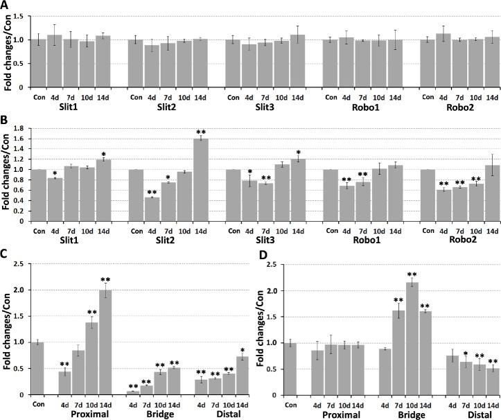
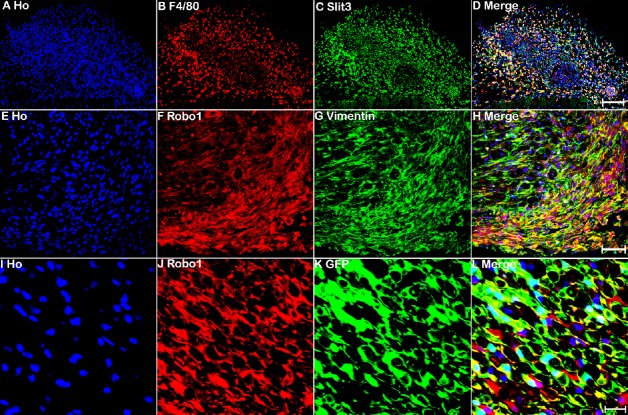
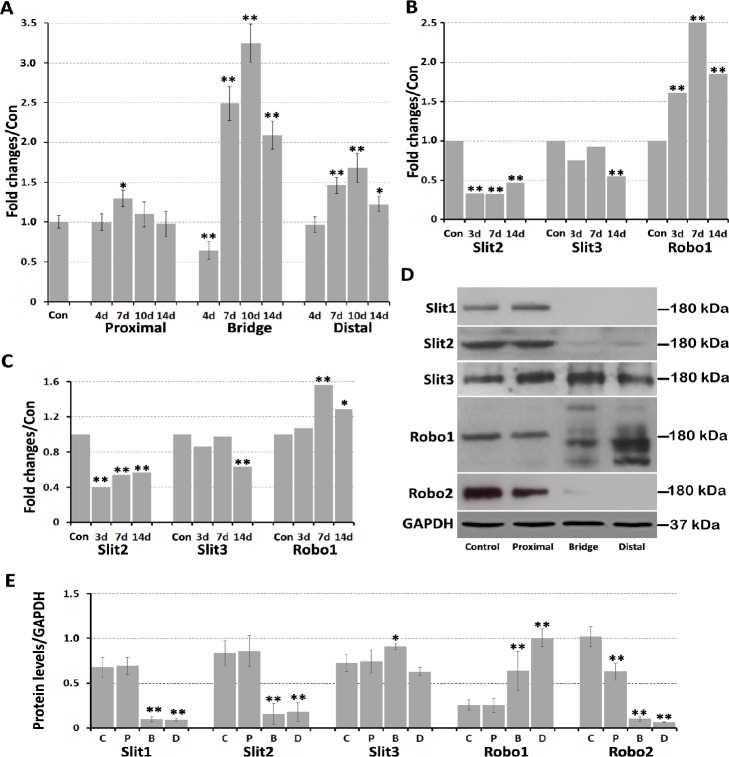
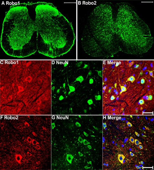
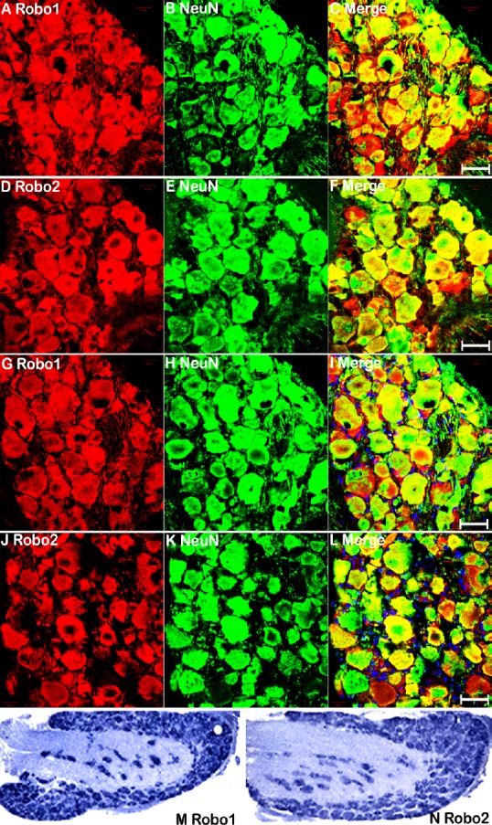
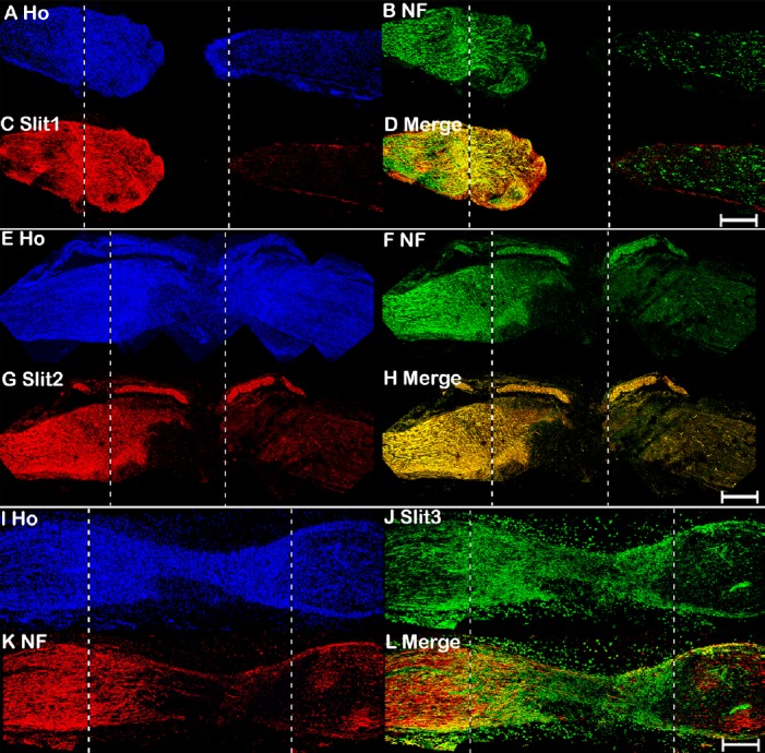
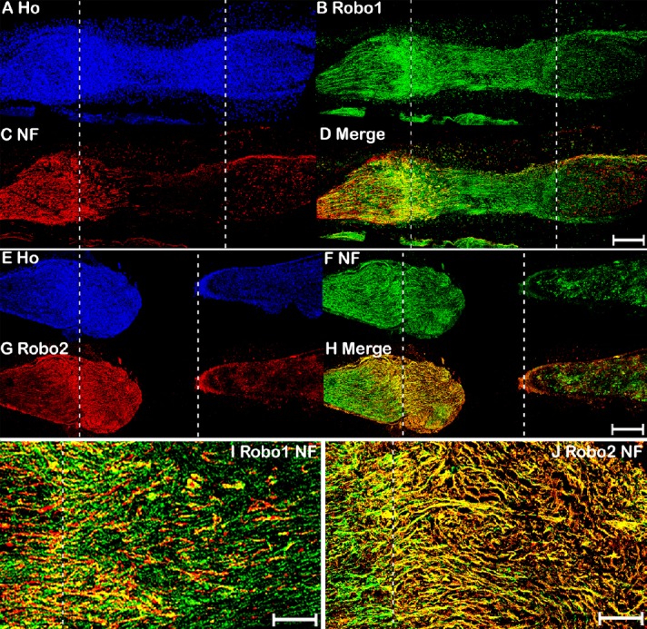
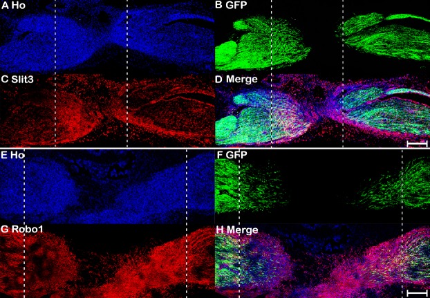
The Slit family of axon guidance cues act as repulsive molecules for precise axon pathfinding and neuronal migration during nervous system development through interactions with specific Robo receptors. Although we previously reported that Slit1-3 and their receptors Robo1 and Robo2 are highly expressed in the adult mouse peripheral nervous system, how this expression changes after injury has not been well studied. Herein, we constructed a peripheral nerve injury mouse model by transecting the right sciatic nerve. At 14 days after injury, quantitative real-time polymerase chain reaction was used to detect mRNA expression of Slit1-3 and Robo1-2 in L4-5 spinal cord and dorsal root ganglia, as well as the sciatic nerve. Immunohistochemical analysis was performed to examine Slit1-3, Robo1-2, neurofilament heavy chain, F4/80, and vimentin in L4-5 spinal cord, L4 dorsal root ganglia, and the sciatic nerve. Co-expression of Slit1-3 and Robo1-2 in L4 dorsal root ganglia was detected by in situ hybridization. In addition, Slit1-3 and Robo1-2 protein expression in L4-5 spinal cord, L4 dorsal root ganglia, and sciatic nerve were detected by western blot assay. The results showed no significant changes of Slit1-3 or Robo1-2 mRNA expression in the spinal cord within 14 days after injury. In the dorsal root ganglion, Slit1-3 and Robo1-2 mRNA expression were initially downregulated within 4 days after injury; however, Robo1-2 mRNA expression returned to the control level, while Slit1-3 mRNA expression remained upregulated during regeneration from 4-14 days after injury. In the sciatic nerve, Slit1-3 and their receptors Robo1-2 were all expressed in the proximal nerve stump; however, Slit1, Slit2, and Robo2 were barely detectable in the nerve bridge and distal nerve stump within 14 days after injury. Slit3 was highly ex-pressed in macrophages surrounding the nerve bridge and slightly downregulated in the distal nerve stump within 14 days after injury. Robo1 was upregulated in vimentin-positive cells and migrating Schwann cells inside the nerve bridge. Robo1 was also upregulated in Schwann cells of the distal nerve stump within 14 days after injury. Our findings indicate that Slit3 is the major ligand expressed in the nerve bridge and distal nerve stump during peripheral nerve regeneration, and Slit3/Robo signaling could play a key role in peripheral nerve repair after injury. This study was approved by Plymouth University Animal Welfare Ethical Review Board (approval No. 30/3203) on April 12, 2014.

摘要

在神经系统发育过程中,轴突导向因子Slit家族作为排斥分子,通过与特定的Robo受体相互作用,实现精确的轴突路径寻找和神经元迁移。尽管我们之前报道过Slit1 - 3及其受体Robo1和Robo2在成年小鼠外周神经系统中高表达,但损伤后这种表达如何变化尚未得到充分研究。在此,我们通过横断右侧坐骨神经构建了外周神经损伤小鼠模型。在损伤后14天,采用定量实时聚合酶链反应检测L4 - 5脊髓、背根神经节以及坐骨神经中Slit1 - 3和Robo1 - 2的mRNA表达。进行免疫组织化学分析,检测L4 - 5脊髓、L4背根神经节和坐骨神经中的Slit1 - 3、Robo1 - 2、神经丝重链、F4/80和波形蛋白。通过原位杂交检测L4背根神经节中Slit1 - 3和Robo1 - 2的共表达。此外,采用蛋白质免疫印迹法检测L4 - 5脊髓、L4背根神经节和坐骨神经中Slit1 - 3和Robo1 - 2蛋白表达。结果显示,损伤后14天内脊髓中Slit1 - 3或Robo1 - 2的mRNA表达无显著变化。在背根神经节中,损伤后4天内Slit1 - 3和Robo1 - 2的mRNA表达最初下调;然而,Robo1 - 2的mRNA表达在损伤后4 - 14天再生过程中恢复到对照水平,而Slit1 - 3的mRNA表达仍上调。在坐骨神经中,Slit1 - 3及其受体Robo1 - 2均在近端神经残端表达;然而,损伤后14天内神经桥和远端神经残端中几乎检测不到Slit1、Slit2和Robo2。Slit3在神经桥周围的巨噬细胞中高表达,损伤后14天内在远端神经残端中略有下调。Robo1在神经桥内波形蛋白阳性细胞和迁移的雪旺细胞中上调。损伤后14天内Robo1在远端神经残端的雪旺细胞中也上调。我们的研究结果表明,Slit3是外周神经再生过程中在神经桥和远端神经残端表达的主要配体,Slit3/Robo信号可能在损伤后外周神经修复中起关键作用。本研究于2014年4月12日获得普利茅斯大学动物福利伦理审查委员会批准(批准号30/3203)。

https://cdn.ncbi.nlm.nih.gov/pmc/blobs/f695/6990781/bdca76ffaebf/NRR-15-948-g009.jpg
https://cdn.ncbi.nlm.nih.gov/pmc/blobs/f695/6990781/b79ff0ee7d4b/NRR-15-948-g002.jpg
https://cdn.ncbi.nlm.nih.gov/pmc/blobs/f695/6990781/b18e6b25ad76/NRR-15-948-g003.jpg
https://cdn.ncbi.nlm.nih.gov/pmc/blobs/f695/6990781/57cfde296389/NRR-15-948-g004.jpg
https://cdn.ncbi.nlm.nih.gov/pmc/blobs/f695/6990781/ea47ea2b3069/NRR-15-948-g005.jpg
https://cdn.ncbi.nlm.nih.gov/pmc/blobs/f695/6990781/a0162fbef9ea/NRR-15-948-g006.jpg
https://cdn.ncbi.nlm.nih.gov/pmc/blobs/f695/6990781/c8d8759dc576/NRR-15-948-g007.jpg
https://cdn.ncbi.nlm.nih.gov/pmc/blobs/f695/6990781/73577d8e0fbd/NRR-15-948-g008.jpg
https://cdn.ncbi.nlm.nih.gov/pmc/blobs/f695/6990781/bdca76ffaebf/NRR-15-948-g009.jpg

相似文献

1
Dynamic expression of Slit1-3 and Robo1-2 in the mouse peripheral nervous system after injury.损伤后小鼠外周神经系统中Slit1-3和Robo1-2的动态表达
Neural Regen Res. 2020 May;15(5):948-958. doi: 10.4103/1673-5374.268930.
2
Expression patterns of Slit and Robo family members in adult mouse spinal cord and peripheral nervous system.成年小鼠脊髓和周围神经系统中Slit和Robo家族成员的表达模式。
PLoS One. 2017 Feb 24;12(2):e0172736. doi: 10.1371/journal.pone.0172736. eCollection 2017.
3
Dynamic changes in Robo2 and Slit1 expression in adult rat dorsal root ganglion and sciatic nerve after peripheral and central axonal injury.成年大鼠外周和中枢轴突损伤后背根神经节和坐骨神经中Robo2和Slit1表达的动态变化
Neurosci Res. 2006 Nov;56(3):314-21. doi: 10.1016/j.neures.2006.07.014. Epub 2006 Sep 18.
4
Expression of the vertebrate Slit gene family and their putative receptors, the Robo genes, in the developing murine kidney.脊椎动物Slit基因家族及其假定受体Robo基因在发育中的小鼠肾脏中的表达。
Mech Dev. 2000 Jun;94(1-2):213-7. doi: 10.1016/s0925-4773(00)00313-0.
5
Macrophage-Derived Slit3 Controls Cell Migration and Axon Pathfinding in the Peripheral Nerve Bridge.巨噬细胞衍生的 Slit3 控制外周神经桥中的细胞迁移和轴突寻迹。
Cell Rep. 2019 Feb 5;26(6):1458-1472.e4. doi: 10.1016/j.celrep.2018.12.081.
6
Classic axon guidance molecules control correct nerve bridge tissue formation and precise axon regeneration.经典的轴突导向分子控制正确的神经桥组织形成和精确的轴突再生。
Neural Regen Res. 2020 Jan;15(1):6-9. doi: 10.4103/1673-5374.264441.
7
Quantification of SLIT-ROBO transcripts in hepatocellular carcinoma reveals two groups of genes with coordinate expression.肝细胞癌中SLIT-ROBO转录本的定量分析揭示了两组具有协同表达的基因。
BMC Cancer. 2008 Dec 29;8:392. doi: 10.1186/1471-2407-8-392.
8
Differential expression of microRNAs in dorsal root ganglia after sciatic nerve injury.背根神经节损伤后 microRNAs 的差异表达。
Neural Regen Res. 2014 May 15;9(10):1031-40. doi: 10.4103/1673-5374.133164.
9
Spatiotemporal expression patterns of slit and robo genes in the rat brain.缝隙基因和轮状蛋白基因在大鼠大脑中的时空表达模式。
J Comp Neurol. 2002 Jan 7;442(2):130-55. doi: 10.1002/cne.10068.
10
Novel regulated expression of the SLIT/ROBO pathway in the ovary: possible role during luteolysis in women.卵巢中SLIT/ROBO通路的新型调控表达:对女性黄体溶解可能具有的作用。
Endocrinology. 2008 Oct;149(10):5024-34. doi: 10.1210/en.2008-0204. Epub 2008 Jun 19.

引用本文的文献

1
The Role of SLIT3-ROBO4 Signaling in Endoplasmic Reticulum Stress-Induced Delayed Corneal Epithelial and Nerve Regeneration.SLIT3-ROBO4信号通路在内质网应激诱导的角膜上皮和神经再生延迟中的作用
Invest Ophthalmol Vis Sci. 2024 May 1;65(5):8. doi: 10.1167/iovs.65.5.8.
2
Mechanism of Robo1 in the pentylenetetrazol-kindled epilepsy mouse model.Robo1在戊四氮点燃癫痫小鼠模型中的作用机制
Ibrain. 2023 Aug 15;9(4):369-380. doi: 10.1002/ibra.12127. eCollection 2023 Winter.
3
N-cadherin directs the collective Schwann cell migration required for nerve regeneration through Slit2/3-mediated contact inhibition of locomotion.

本文引用的文献

1
Macrophage-Derived Slit3 Controls Cell Migration and Axon Pathfinding in the Peripheral Nerve Bridge.巨噬细胞衍生的 Slit3 控制外周神经桥中的细胞迁移和轴突寻迹。
Cell Rep. 2019 Feb 5;26(6):1458-1472.e4. doi: 10.1016/j.celrep.2018.12.081.
2
Repair of peripheral nerve defects by nerve transposition using small gap bio-sleeve suture with different inner diameters at both ends.使用两端内径不同的小间隙生物套管缝合技术进行神经转位修复周围神经缺损。
Neural Regen Res. 2019 Apr;14(4):706-712. doi: 10.4103/1673-5374.247475.
3
Transection and Crush Models of Nerve Injury to Measure Repair and Remyelination in Peripheral Nerve.
N-钙黏蛋白通过 Slit2/3 介导的运动接触抑制来指导 Schwann 细胞的集体迁移,这对于神经再生是必需的。
Elife. 2024 Apr 9;13:e88872. doi: 10.7554/eLife.88872.
4
The histomorphological and stereological assessment of rat dorsal root ganglion tissues after various types of sciatic nerve injury.大鼠各种类型坐骨神经损伤后背根神经节组织的组织形态学和体视学评估。
Histochem Cell Biol. 2024 Feb;161(2):145-163. doi: 10.1007/s00418-023-02242-0. Epub 2023 Oct 19.
5
Transcriptomic and Proteomic of Gastrocnemius Muscle in Peripheral Artery Disease.腓肠肌在周围动脉疾病中的转录组学和蛋白质组学研究。
Circ Res. 2023 May 26;132(11):1428-1443. doi: 10.1161/CIRCRESAHA.122.322325. Epub 2023 May 8.
6
Single-cell transcriptomic analysis reveals the adverse effects of cadmium on the trajectory of neuronal maturation.单细胞转录组分析揭示了镉对神经元成熟轨迹的不良影响。
Cell Biol Toxicol. 2023 Aug;39(4):1697-1713. doi: 10.1007/s10565-022-09775-5. Epub 2022 Sep 17.
7
Magnetic Resonance Imaging Investigation of Neuroplasticity After Ischemic Stroke in Tetramethylpyrazine-Treated Rats.川芎嗪治疗大鼠缺血性中风后神经可塑性的磁共振成像研究
Front Pharmacol. 2022 Apr 26;13:851746. doi: 10.3389/fphar.2022.851746. eCollection 2022.
8
Will Cannabigerol Trigger Neuroregeneration after a Spinal Cord Injury? An In Vitro Answer from NSC-34 Scratch-Injured Cells Transcriptome.大麻二酚会引发脊髓损伤后的神经再生吗?来自NSC - 34划痕损伤细胞转录组的体外研究答案。
Pharmaceuticals (Basel). 2022 Jan 19;15(2):117. doi: 10.3390/ph15020117.
9
Role of Slit/Robo Signaling pathway in Bone Metabolism.Slit/Robo 信号通路在骨代谢中的作用。
Int J Biol Sci. 2022 Jan 9;18(3):1303-1312. doi: 10.7150/ijbs.66931. eCollection 2022.
10
Pericyte‑derived extracellular vesicles‑mimetic nanovesicles improves peripheral nerve regeneration in mouse models of sciatic nerve transection.外泌体模拟纳米囊泡促进坐骨神经横断小鼠模型的周围神经再生
Int J Mol Med. 2022 Feb;49(2). doi: 10.3892/ijmm.2021.5073. Epub 2021 Dec 22.
用于测量周围神经修复和髓鞘再生的神经损伤横断和挤压模型
Methods Mol Biol. 2018;1791:251-262. doi: 10.1007/978-1-4939-7862-5_20.
4
Expression patterns of Slit and Robo family members in adult mouse spinal cord and peripheral nervous system.成年小鼠脊髓和周围神经系统中Slit和Robo家族成员的表达模式。
PLoS One. 2017 Feb 24;12(2):e0172736. doi: 10.1371/journal.pone.0172736. eCollection 2017.
5
Slit-Robo signaling.Slit-Robo信号传导
Development. 2016 Sep 1;143(17):3037-44. doi: 10.1242/dev.132829.
6
Gene expression analysis at multiple time-points identifies key genes for nerve regeneration.多个时间点的基因表达分析确定了神经再生的关键基因。
Muscle Nerve. 2017 Mar;55(3):373-383. doi: 10.1002/mus.25225. Epub 2016 Nov 23.
7
The lh3 Glycosyltransferase Directs Target-Selective Peripheral Nerve Regeneration.LH3糖基转移酶指导靶向选择性周围神经再生。
Neuron. 2015 Nov 18;88(4):691-703. doi: 10.1016/j.neuron.2015.10.004. Epub 2015 Nov 5.
8
Slit and Receptor Tyrosine Phosphatase 69D Confer Spatial Specificity to Axon Branching via Dscam1.Slit和受体酪氨酸磷酸酶69D通过Dscam1赋予轴突分支空间特异性。
Cell. 2015 Aug 27;162(5):1140-54. doi: 10.1016/j.cell.2015.08.003.
9
Macrophage-Induced Blood Vessels Guide Schwann Cell-Mediated Regeneration of Peripheral Nerves.巨噬细胞诱导的血管引导雪旺细胞介导的周围神经再生。
Cell. 2015 Aug 27;162(5):1127-39. doi: 10.1016/j.cell.2015.07.021. Epub 2015 Aug 13.
10
Visualizing peripheral nerve regeneration by whole mount staining.通过整体染色观察周围神经再生
PLoS One. 2015 Mar 4;10(3):e0119168. doi: 10.1371/journal.pone.0119168. eCollection 2015.