Department of Chemistry, Purdue University, West Lafayette, IN, USA.
Biological Sciences Division, Pacific Northwest National Laboratory, Richland, WA, USA.
Nat Protoc. 2019 Dec;14(12):3445-3470. doi: 10.1038/s41596-019-0237-4. Epub 2019 Nov 13.
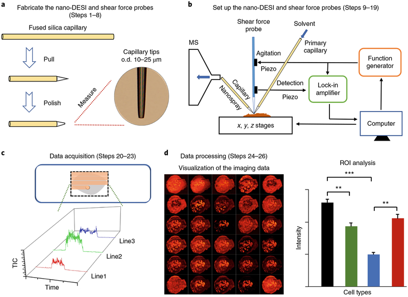
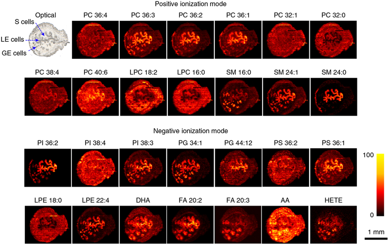
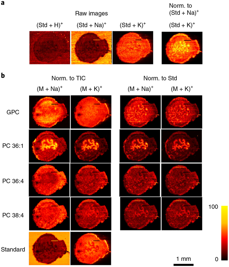
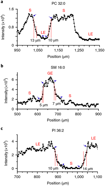
Mass spectrometry imaging (MSI) enables label-free spatial mapping of hundreds of biomolecules in tissue sections. This capability provides valuable information on tissue heterogeneity that is difficult to obtain using population-averaged assays. Despite substantial developments in both instrumentation and methodology, MSI of tissue samples at single-cell resolution remains challenging. Herein, we describe a protocol for robust imaging of tissue sections with a high (better than 10-μm) spatial resolution using nanospray desorption electrospray ionization (nano-DESI) mass spectrometry, an ambient ionization technique that does not require sample pretreatment before analysis. In this protocol, mouse uterine tissue is used as a model system to illustrate both the workflow and data obtained in these experiments. We provide a detailed description of the nano-DESI MSI platform, fabrication of the nano-DESI and shear force probes, shear force microscopy experiments, spectral acquisition, and data processing. A properly trained researcher (e.g., technician, graduate student, or postdoc) can complete all the steps from probe fabrication to data acquisition and processing within a single day. We also describe a new strategy for acquiring both positive- and negative-mode imaging data in the same experiment. This is achieved by alternating between positive and negative data acquisition modes during consecutive line scans. Using our imaging approach, hundreds of high-quality ion images were obtained from a single uterine section. This protocol enables sensitive and quantitative imaging of lipids and metabolites in heterogeneous tissue sections with high spatial resolution, which is critical to understanding biochemical processes occurring in biological tissues.
质谱成像(MSI)能够在组织切片中无标记地对数百种生物分子进行空间定位。这种能力提供了有关组织异质性的有价值的信息,而这些信息很难通过平均人群测定获得。尽管在仪器和方法学方面都取得了实质性的发展,但单细胞分辨率的组织样本 MSI 仍然具有挑战性。在此,我们描述了一种使用纳米喷雾解吸电喷雾电离(nano-DESI)质谱进行高(优于 10-μm)空间分辨率的组织切片稳健成像的方案,这是一种无需在分析前进行样品预处理的环境电离技术。在本方案中,我们以小鼠子宫组织为模型系统,说明了这些实验中的工作流程和获得的数据。我们详细描述了 nano-DESI MSI 平台、nano-DESI 和剪切力探针的制造、剪切力显微镜实验、光谱采集和数据处理。经过适当培训的研究人员(例如技术人员、研究生或博士后)可以在一天内完成从探针制造到数据采集和处理的所有步骤。我们还描述了一种在同一个实验中同时获取正离子和负离子成像数据的新策略。这是通过在连续线扫描过程中在正离子和负离子数据采集模式之间交替来实现的。使用我们的成像方法,从单个子宫切片中获得了数百张高质量的离子图像。该方案能够以高空间分辨率对异质组织切片中的脂质和代谢物进行灵敏且定量的成像,这对于理解生物组织中发生的生化过程至关重要。