Harvard-MIT Division of Health Sciences and Technology, Harvard Medical School and Massachusetts Institute of Technology, Cambridge, MA, USA.
Department of Electrical Engineering and Computer Science and Research Laboratory of Electronics, Massachusetts Institute of Technology, Cambridge, MA, USA.
Mod Pathol. 2020 May;33(5):916-923. doi: 10.1038/s41379-019-0408-4. Epub 2019 Nov 19.
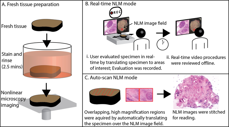
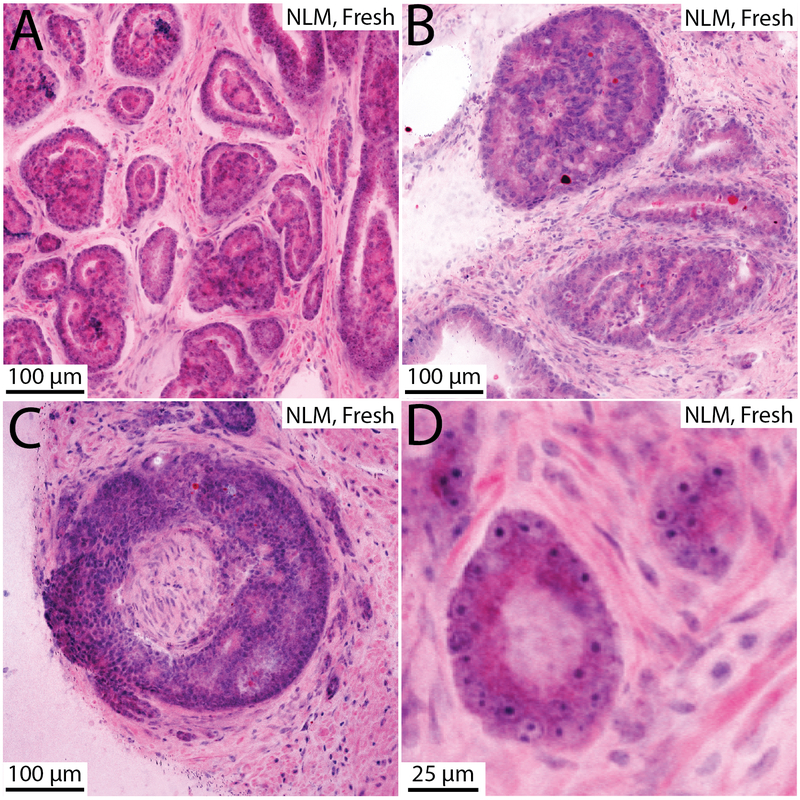
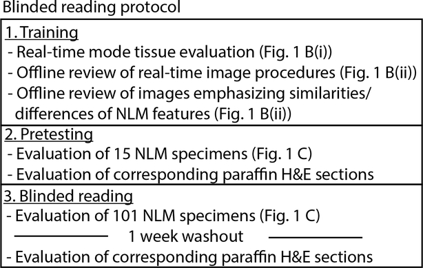
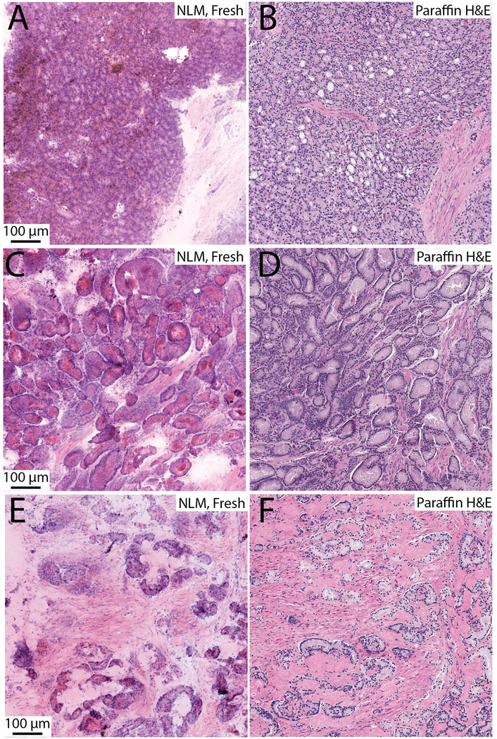
Intraoperative evaluation of specimens during radical prostatectomy using frozen sections can be time and labor intensive. Nonlinear microscopy (NLM) is a fluorescence microscopy technique that can rapidly generate images that closely resemble H&E histology in freshly excised tissue, without requiring freezing or microtome sectioning. Specimens are stained with nuclear and cytoplasmic/stromal fluorophores, and NLM evaluation can begin within 3 min of grossing. Fluorescence signals can be displayed using an H&E color scale, facilitating pathologist interpretation. This study evaluates the accuracy of prostate cancer detection in a blinded reading of NLM images compared with the gold standard of formalin-fixed, paraffin-embedded H&E histology. A total of 122 freshly excised prostate specimens were obtained from 40 patients undergoing radical prostatectomy. The prostates were grossed, dissected into specimens of ~10 × 10 mm with 1-4 mm thickness, stained for 2 min for nuclear and cytoplasmic/stromal contrast, and then rinsed with saline for 30 s. NLM images were acquired and multiple images were stitched together to generate large field of view, centimeter-scale digital images suitable for reading. Specimens were then processed for standard paraffin H&E. The study protocol consisted of training, pretesting, and blinded reading phases. After a washout period, pathologists read corresponding paraffin H&E slides. Three pathologists achieved a 95% or greater sensitivity with 100% specificity for detecting cancer on NLM compared with paraffin H&E. Pooled sensitivity and specificity was 97.3% (93.7-99.1%; 95% confidence interval) and 100.0% (97.0-100.0%), respectively. Interobserver agreement for NLM reading had a Fleiss κ = 0.95. The high cancer detection accuracy and rapid specimen preparation suggest that NLM may be useful for intraoperative evaluation in radical prostatectomy.
在根治性前列腺切除术期间使用冰冻切片对标本进行术中评估既耗时又费力。非线性显微镜(NLM)是一种荧光显微镜技术,它可以快速生成与新鲜切除组织的 H&E 组织学非常相似的图像,而无需冷冻或切片。标本用核和细胞质/基质荧光染料染色,NLM 评估可以在大体检查后 3 分钟内开始。荧光信号可以使用 H&E 颜色标度显示,便于病理学家进行解释。本研究评估了在与福尔马林固定、石蜡包埋 H&E 组织学的金标准进行盲法阅读相比,NLM 图像在前列腺癌检测中的准确性。总共从 40 名接受根治性前列腺切除术的患者中获得了 122 个新鲜切除的前列腺标本。对前列腺进行大体检查,将其切成~10×10mm 的标本,厚度为 1-4mm,用核和细胞质/基质对比染料染色 2 分钟,然后用生理盐水冲洗 30 秒。获取 NLM 图像,并将多个图像拼接在一起,生成适合阅读的大视野、厘米级数字图像。然后将标本进行标准石蜡 H&E 处理。研究方案包括培训、预测试和盲法阅读阶段。经过洗脱期后,病理学家阅读相应的石蜡 H&E 载玻片。与石蜡 H&E 相比,三名病理学家在 NLM 上检测癌症的灵敏度达到 95%或更高,特异性为 100%。汇总的敏感性和特异性分别为 97.3%(93.7-99.1%;95%置信区间)和 100.0%(97.0-100.0%)。NLM 阅读的观察者间一致性 Fleiss κ 值为 0.95。高癌症检测准确性和快速标本制备表明,NLM 可能对根治性前列腺切除术的术中评估有用。