Department of Radiology, Fujian Maternity and Child Health Hospital, Fuzhou, Fujian, China.
Department of Radiology, Fujian Medical University Union Hospital, Fuzhou, Fujian, China.
BMC Med Imaging. 2020 Nov 25;20(1):125. doi: 10.1186/s12880-020-00525-9.
Reported date of last menstrual period and ultrasonography measurements are the most commonly used methods for determining gestational age in antenatal life. However, the mother cannot always determine the last menstrual period with certainty, and ultrasonography measurements are accurate only in the first trimester. We aimed to assess the ability of various biometric measurements on magnetic resonance imaging (MRI) in determining the accurate gestational age of an individual fetus in the second half of gestation.
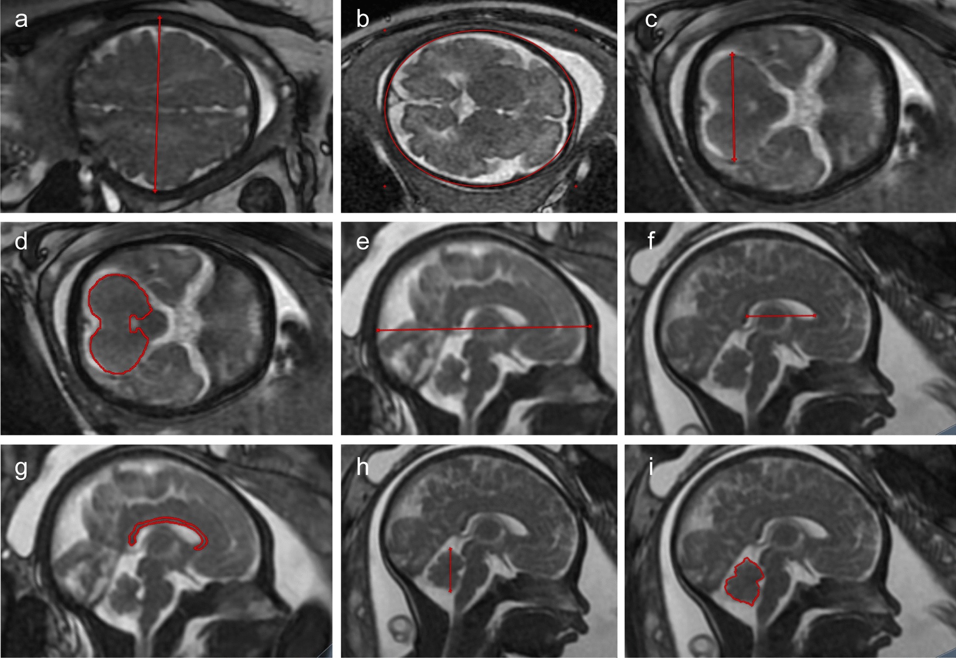
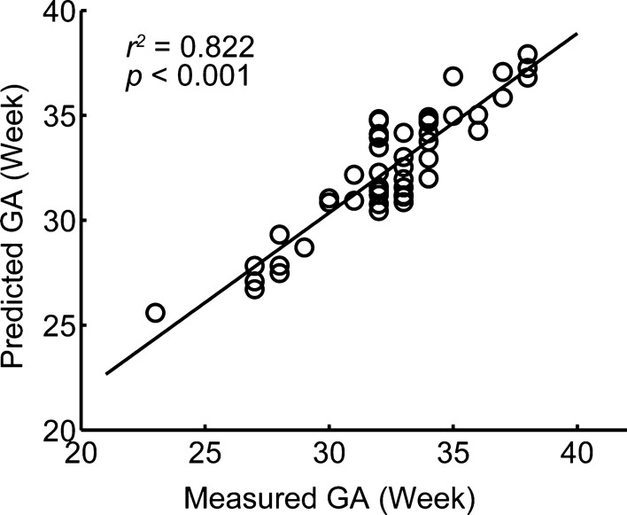
We used MRI to scan a total of 637 fetuses ranging in age from 22 to 40 gestational weeks. We evaluated 9 standard fetal 2D biometric parameters, and regression models were fitted to assess normal fetal brain development. A stepwise linear regression model was constructed to predict gestational age, and measurement accuracy was determined in a held-out, unseen test sample (n = 49).
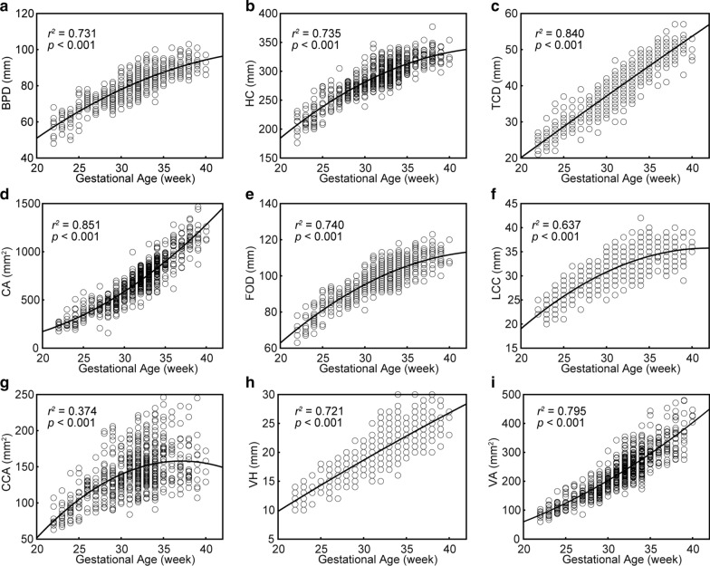
A second-order polynomial regression model was found to be the best descriptor of biometric measures including brain bi-parietal diameter, head circumference, and fronto-occipital diameter in relation to normal fetal growth. Normal fetuses showed divergent growth patterns for the cerebrum and cerebellum, where the cerebrum undergoes rapid growth in the second trimester, while the cerebellum undergoes rapid growth in the third trimester. Moreover, a linear model based on biometrics of brain bi-parietal diameter, length of the corpus callosum, vermis area, transverse cerebellar diameter, and cerebellar area accurately predicted gestational age in the second and third trimesters (cross-validation R = 0.822, p < 0.001).
These results support the use of MRI biometry charts to improve MRI evaluation of fetal growth and suggest that MRI biometry measurements offer a potential estimation model of fetal gestational age in the second half of gestation, which is vital to any assessment of pregnancy, fetal development, and neonatal care.
末次月经日期和超声测量是产前确定胎龄最常用的方法。然而,母亲并不总是能确定末次月经日期,并且超声测量仅在孕早期准确。我们旨在评估磁共振成像(MRI)中各种生物测量值在确定个体胎儿妊娠后半期准确胎龄方面的能力。
我们使用 MRI 扫描了总共 637 个年龄在 22 至 40 孕周的胎儿。我们评估了 9 个标准的胎儿 2D 生物测量参数,并拟合回归模型以评估正常胎儿脑发育。构建了一个逐步线性回归模型来预测胎龄,并在未见过的测试样本(n=49)中确定测量精度。
发现二次多项式回归模型是描述与正常胎儿生长相关的生物测量值(包括大脑双额径、头围和额枕径)的最佳描述符。正常胎儿的大脑和小脑呈现出不同的生长模式,其中大脑在孕中期快速生长,而小脑在孕晚期快速生长。此外,基于大脑双额径、胼胝体长度、蚓部面积、横径小脑和小脑面积的生物测量值的线性模型准确预测了第二和第三孕期的胎龄(交叉验证 R=0.822,p<0.001)。
这些结果支持使用 MRI 生物测量图来改善 MRI 对胎儿生长的评估,并表明 MRI 生物测量值为妊娠后半期胎儿胎龄提供了潜在的估计模型,这对任何妊娠、胎儿发育和新生儿护理的评估都至关重要。