• 文献检索
  • 文档翻译
  • 深度研究
  • 学术资讯
  • Suppr Zotero 插件Zotero 插件
  • 邀请有礼
  • 套餐&价格
  • 历史记录
应用&插件
Suppr Zotero 插件Zotero 插件浏览器插件Mac 客户端Windows 客户端微信小程序
定价
高级版会员购买积分包购买API积分包
服务
文献检索文档翻译深度研究API 文档MCP 服务
关于我们
关于 Suppr公司介绍联系我们用户协议隐私条款
关注我们

Suppr 超能文献

核心技术专利:CN118964589B侵权必究
粤ICP备2023148730 号-1Suppr @ 2026

文献检索

告别复杂PubMed语法,用中文像聊天一样搜索,搜遍4000万医学文献。AI智能推荐,让科研检索更轻松。

立即免费搜索

文件翻译

保留排版,准确专业,支持PDF/Word/PPT等文件格式,支持 12+语言互译。

免费翻译文档

深度研究

AI帮你快速写综述,25分钟生成高质量综述,智能提取关键信息,辅助科研写作。

立即免费体验

HCC患者和结直肠癌肝转移患者中EOB-GD-DTPA和Gd-BT-DO3A磁共振对比研究的定性评估

Qualitative assessment of EOB-GD-DTPA and Gd-BT-DO3A MR contrast studies in HCC patients and colorectal liver metastases.

作者信息

Granata Vincenza, Fusco Roberta, Maio Francesca, Avallone Antonio, Nasti Guglielmo, Palaia Raffaele, Albino Vittorio, Grassi Roberto, Izzo Francesco, Petrillo Antonella

机构信息

Division of Radiology, ISTITUTO NAZIONALE TUMORI - IRCCS - FONDAZIONE G. PASCALE, NAPOLI, ITALIA, Naples, Italy.

2Division of Radiology, Università degli Studi di Napoli Federico II, Naples, Italy.

出版信息

Infect Agent Cancer. 2019 Nov 27;14:40. doi: 10.1186/s13027-019-0264-3. eCollection 2019.

DOI:10.1186/s13027-019-0264-3
PMID:31798677
原文链接:https://pmc.ncbi.nlm.nih.gov/articles/PMC6882051/
Abstract

AIM

To compare liver-specific EOB-GD-DTPA and liver-non-specific Gd-BT-DO3A MR, in hepatocellular carcinoma (HCC) and liver colorectal metastases.

MATERIAL AND METHODS

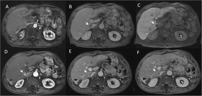
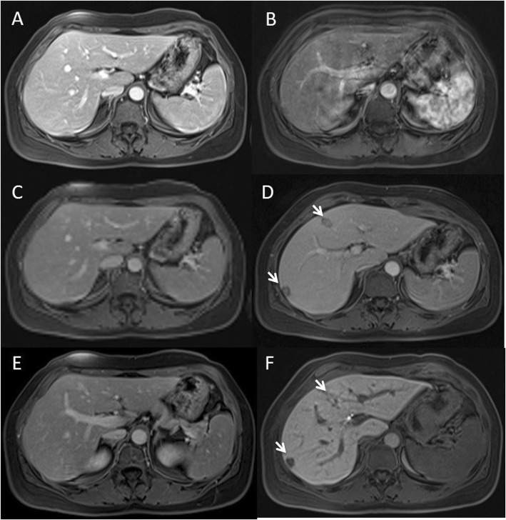
Seventy HCC patients with 158 nodules and 90 colorectal liver metastases (mCRC) with 370 lesions were included in the retrospective analysis. HCC patients underwent MR at 0 time (MR0), after 3 (MR3) and 6 months (MR6) using two different CM; 69 mCRC patients underwent MR with Gd-EOB-BTPA and 21 mCRC patients with Gd-BT-DO3A. We evaluated arterial phase hyperenhancement, lesion-to-liver contrast during portal phase, hepatobiliary phase parenchymal hyperenhancement.

RESULTS

In HCC patients arterial phase hyperenhancement degree was statistically higher ( = 0.03) with Gd-BT-DO3A (mean 4) than GD-EOB-DTPA (mean 2.6), while we found no significant statistical differences among mean (2.6) values at MR0 and MR6 using GD-EOB-DTPA. For all 209 patients underwent Gd-EOB-DTPA, we found that lesion-to-liver contrast during portal phase mean value was 4 while for patients underwent MR with Gd-BT-DO3A was 3 ( = 0.04). For HCC hepatobiliary phase parenchymal hyperenhancement mean value was 2.4. For mCRC patients: among 63 patients underwent previous chemotherapy hepatobiliary phase parenchymal hyperenhancement mean value was 3.1 while for 6 patients no underwent previous chemotherapy was 4 ( = 0.05).

CONCLUSIONS

Gd-EOB-DTPA should be chosen in pre surgical setting in patients with colorectal liver metastases.

摘要

目的

比较肝细胞癌(HCC)和肝结肠转移瘤中肝脏特异性EOB - GD - DTPA和肝脏非特异性Gd - BT - DO3A磁共振成像(MR)。

材料与方法

回顾性分析纳入70例患有158个结节的HCC患者和90例患有370个病灶的结肠肝转移瘤(mCRC)患者。HCC患者在0时(MR0)、3个月后(MR3)和6个月时(MR6)使用两种不同的对比剂进行MR检查;69例mCRC患者使用Gd - EOB - BTPA进行MR检查,21例mCRC患者使用Gd - BT - DO3A进行MR检查。我们评估了动脉期高增强、门静脉期病灶与肝脏的对比、肝胆期实质高增强情况。

结果

在HCC患者中,使用Gd - BT - DO3A时动脉期高增强程度在统计学上更高(P = 0.03)(平均值为4),高于GD - EOB - DTPA(平均值为2.6),而在使用GD - EOB - DTPA时,我们发现在MR0和MR6时平均值(2.6)之间无显著统计学差异。对于所有209例接受Gd - EOB - DTPA检查的患者,我们发现门静脉期病灶与肝脏对比的平均值为4,而对于接受Gd - BT - DO3A MR检查的患者为3(P = 0.04)。对于HCC,肝胆期实质高增强平均值为2.4。对于mCRC患者:在63例先前接受过化疗的患者中,肝胆期实质高增强平均值为3.1,而在6例未接受过先前化疗的患者中为4(P = 0.05)。

结论

对于结肠肝转移瘤患者,在术前应选择Gd - EOB - DTPA。

https://cdn.ncbi.nlm.nih.gov/pmc/blobs/e5ca/6882051/f098d9cb7352/13027_2019_264_Fig2_HTML.jpg
https://cdn.ncbi.nlm.nih.gov/pmc/blobs/e5ca/6882051/2e07fd3a63e6/13027_2019_264_Fig1_HTML.jpg
https://cdn.ncbi.nlm.nih.gov/pmc/blobs/e5ca/6882051/f098d9cb7352/13027_2019_264_Fig2_HTML.jpg
https://cdn.ncbi.nlm.nih.gov/pmc/blobs/e5ca/6882051/2e07fd3a63e6/13027_2019_264_Fig1_HTML.jpg
https://cdn.ncbi.nlm.nih.gov/pmc/blobs/e5ca/6882051/f098d9cb7352/13027_2019_264_Fig2_HTML.jpg

相似文献

1
Qualitative assessment of EOB-GD-DTPA and Gd-BT-DO3A MR contrast studies in HCC patients and colorectal liver metastases.HCC患者和结直肠癌肝转移患者中EOB-GD-DTPA和Gd-BT-DO3A磁共振对比研究的定性评估
Infect Agent Cancer. 2019 Nov 27;14:40. doi: 10.1186/s13027-019-0264-3. eCollection 2019.
2
Detection of pancreatic ductal adenocarcinoma and liver metastases: comparison of Gd-EOB-DTPA-enhanced MR imaging vs. extracellular contrast materials.检测胰腺导管腺癌和肝转移:钆塞酸二钠增强磁共振成像与细胞外对比剂的比较。
Abdom Radiol (NY). 2020 Aug;45(8):2459-2468. doi: 10.1007/s00261-020-02511-9.
3
Impact of gadoxetic acid (Gd-EOB-DTPA)-enhanced magnetic resonance on the non-invasive diagnosis of small hepatocellular carcinoma: a prospective study.钆塞酸二钠(Gd-EOB-DTPA)增强磁共振对小肝细胞癌非侵入性诊断的影响:一项前瞻性研究。
Aliment Pharmacol Ther. 2013 Feb;37(3):355-63. doi: 10.1111/apt.12166. Epub 2012 Dec 2.
4
Intra-individual comparison of hepatocellular carcinoma imaging features on contrast-enhanced computed tomography, gadopentetate dimeglumine-enhanced MRI, and gadoxetic acid-enhanced MRI.肝细胞癌在对比增强计算机断层扫描、钆喷酸葡胺增强磁共振成像和钆塞酸二钠增强磁共振成像上的个体内影像特征比较
Acta Radiol. 2018 Jun;59(6):639-648. doi: 10.1177/0284185117728534. Epub 2017 Aug 20.
5
Imaging Features of Hepatocellular Carcinoma: Quantitative and Qualitative Comparison Between MRI-Enhanced With Gd-EOB-DTPA and Gd-DTPA.肝细胞癌的影像学特征:钆塞酸二钠(Gd-EOB-DTPA)增强 MRI 与 Gd-DTPA 的定量和定性比较
Invest Radiol. 2019 Aug;54(8):494-499. doi: 10.1097/RLI.0000000000000562.
6
Focal hepatic lesions in Gd-EOB-DTPA enhanced MRI: the atlas.钆塞酸二钠增强 MRI 中的局灶性肝脏病变:图谱。
Insights Imaging. 2012 Oct;3(5):451-74. doi: 10.1007/s13244-012-0179-7. Epub 2012 Jun 15.
7
Evaluation of combined Gd-EOB-DTPA and gadobutrol magnetic resonance imaging for the prediction of hepatocellular carcinoma grading.钆塞酸二钠与钆布醇联合磁共振成像用于预测肝细胞癌分级的评估
Acta Radiol. 2016 Aug;57(8):932-8. doi: 10.1177/0284185115616293. Epub 2015 Nov 18.
8
The target sign in colorectal liver metastases: an atypical Gd-EOB-DTPA "uptake" on the hepatobiliary phase of MR imaging.结直肠癌肝转移中的靶征:磁共振成像肝胆期非典型钆塞酸二钠“摄取”
Abdom Imaging. 2015 Oct;40(7):2364-71. doi: 10.1007/s00261-015-0488-7.
9
Therapy response assessment after radioembolization of patients with hepatocellular carcinoma--comparison of MR imaging with gadolinium ethoxybenzyl diethylenetriamine penta-acetic acid and gadobutrol.肝细胞癌患者放射性栓塞术后的治疗反应评估——钆乙氧基苄基二乙三胺五乙酸磁共振成像与钆布醇的比较
J Vasc Interv Radiol. 2015 Jul;26(7):972-9. doi: 10.1016/j.jvir.2015.03.009. Epub 2015 Apr 27.
10
Gd-EOB-DTPA-enhanced magnetic resonance imaging features of hepatic hemangioma compared with enhanced computed tomography.钆塞酸二钠增强磁共振成像与增强 CT 对肝血管瘤的影像学特征比较。
World J Gastroenterol. 2012 Nov 21;18(43):6269-76. doi: 10.3748/wjg.v18.i43.6269.

引用本文的文献

1
An Informative Review of Radiomics Studies on Cancer Imaging: The Main Findings, Challenges and Limitations of the Methodologies.基于癌症影像学的放射组学研究的综述:方法的主要发现、挑战和局限性。
Curr Oncol. 2024 Jan 10;31(1):403-424. doi: 10.3390/curroncol31010027.
2
Colorectal liver metastases patients prognostic assessment: prospects and limits of radiomics and radiogenomics.结直肠癌肝转移患者的预后评估:影像组学和影像基因组学的前景与局限
Infect Agent Cancer. 2023 Mar 16;18(1):18. doi: 10.1186/s13027-023-00495-x.
3
Risk Assessment and Cholangiocarcinoma: Diagnostic Management and Artificial Intelligence.

本文引用的文献

1
Comment on "State of the art in magnetic resonance imaging of hepatocellular carcinoma": the role of DWI.关于《肝细胞癌磁共振成像的现状》的评论:扩散加权成像的作用
Radiol Oncol. 2019 Jul 13;53(3):369-370. doi: 10.2478/raon-2019-0031.
2
Radiofrequency Ablation and Microwave Ablation in Liver Tumors: An Update.射频消融与微波消融在肝脏肿瘤治疗中的应用进展
Oncologist. 2019 Oct;24(10):e990-e1005. doi: 10.1634/theoncologist.2018-0337. Epub 2019 Jun 19.
3
Microvascular invasion and grading in hepatocellular carcinoma: correlation with major and ancillary features according to LIRADS.
风险评估与胆管癌:诊断管理与人工智能
Biology (Basel). 2023 Jan 29;12(2):213. doi: 10.3390/biology12020213.
4
Diagnostic Management of Gastroenteropancreatic Neuroendocrine Neoplasms: Technique Optimization and Tips and Tricks for Radiologists.胃肠胰神经内分泌肿瘤的诊断管理:放射科医师的技术优化及技巧。
Tomography. 2023 Jan 27;9(1):217-246. doi: 10.3390/tomography9010018.
5
Imaging Features of Main Hepatic Resections: The Radiologist Challenging.肝主要切除术的影像学特征:放射科医生面临的挑战。
J Pers Med. 2023 Jan 10;13(1):134. doi: 10.3390/jpm13010134.
6
Risk Assessment and Pancreatic Cancer: Diagnostic Management and Artificial Intelligence.风险评估与胰腺癌:诊断管理与人工智能
Cancers (Basel). 2023 Jan 5;15(2):351. doi: 10.3390/cancers15020351.
7
Machine Learning Approaches with Textural Features to Calculate Breast Density on Mammography.基于纹理特征的机器学习方法计算乳腺 X 线摄影中的乳腺密度。
Curr Oncol. 2023 Jan 7;30(1):839-853. doi: 10.3390/curroncol30010064.
8
Multimodality Imaging Assessment of Desmoid Tumors: The Great Mime in the Era of Multidisciplinary Teams.韧带样瘤的多模态成像评估:多学科团队时代的“伟大模仿者”
J Pers Med. 2022 Jul 16;12(7):1153. doi: 10.3390/jpm12071153.
9
A Narrative Review on LI-RADS Algorithm in Liver Tumors: Prospects and Pitfalls.关于肝脏肿瘤中LI-RADS算法的叙述性综述:前景与陷阱
Diagnostics (Basel). 2022 Jul 7;12(7):1655. doi: 10.3390/diagnostics12071655.
10
Diffusion weighted imaging and diffusion kurtosis imaging in abdominal oncological setting: why and when.腹部肿瘤学中的扩散加权成像和扩散峰度成像:为何及何时应用。
Infect Agent Cancer. 2022 Jun 9;17(1):25. doi: 10.1186/s13027-022-00441-3.
肝细胞癌的微血管侵犯和分级:根据 LIRADS 与主要和辅助特征的相关性。
Abdom Radiol (NY). 2019 Aug;44(8):2788-2800. doi: 10.1007/s00261-019-02056-6.
4
A radiologist's point of view in the presurgical and intraoperative setting of colorectal liver metastases.结直肠肝转移术前和术中的放射科医师观点。
Future Oncol. 2018 Sep;14(21):2189-2206. doi: 10.2217/fon-2018-0080. Epub 2018 Aug 7.
5
The current role and future prospectives of functional parameters by diffusion weighted imaging in the assessment of histologic grade of HCC.扩散加权成像功能参数在肝癌组织学分级评估中的当前作用及未来展望。
Infect Agent Cancer. 2018 Jul 3;13:23. doi: 10.1186/s13027-018-0194-5. eCollection 2018.
6
Evolution of indeterminate hepatocellular nodules at Gd-EOB-DPTA-enhanced MRI in cirrhotic patients.肝硬化患者 Gd-EOB-DPTA 增强 MRI 下不定型肝细胞结节的演变。
Radiol Med. 2018 Jul;123(7):489-497. doi: 10.1007/s11547-018-0873-8. Epub 2018 Mar 15.
7
Gadoxetate disodium-induced tachypnoea and the effect of dilution method: a proof-of-concept study in mice.钆塞酸二钠引起的呼吸急促及稀释法的作用:在小鼠中的概念验证研究。
Eur Radiol. 2018 Feb;28(2):692-697. doi: 10.1007/s00330-017-5037-z. Epub 2017 Sep 11.
8
Critical analysis of the major and ancillary imaging features of LI-RADS on 127 proven HCCs evaluated with functional and morphological MRI: Lights and shadows.对通过功能和形态学MRI评估的127例经证实的肝细胞癌(HCC)的LI-RADS主要和辅助影像学特征进行批判性分析:亮点与阴影。
Oncotarget. 2017 Apr 19;8(31):51224-51237. doi: 10.18632/oncotarget.17227. eCollection 2017 Aug 1.
9
Uncommon neoplasms of the biliary tract: radiological findings.胆道罕见肿瘤:影像学表现
Br J Radiol. 2017 Oct;90(1078):20160561. doi: 10.1259/bjr.20160561. Epub 2017 Jul 21.
10
Diagnostic accuracy of magnetic resonance, computed tomography and contrast enhanced ultrasound in radiological multimodality assessment of peribiliary liver metastases.磁共振成像、计算机断层扫描及对比增强超声在肝门周围肝转移瘤放射学多模态评估中的诊断准确性
PLoS One. 2017 Jun 20;12(6):e0179951. doi: 10.1371/journal.pone.0179951. eCollection 2017.