Simonetti Igino, Bruno Federico, Fusco Roberta, Cutolo Carmen, Setola Sergio Venanzio, Patrone Renato, Masciocchi Carlo, Palumbo Pierpaolo, Arrigoni Francesco, Picone Carmine, Belli Andrea, Grassi Roberta, Grassi Francesca, Barile Antonio, Izzo Francesco, Petrillo Antonella, Granata Vincenza
Division of Radiology, Istituto Nazionale Tumori IRCCS Fondazione Pascale-IRCCS di Napoli, 80131 Naples, Italy.
Department of Applied Clinical Sciences and Biotechnology, University of L'Aquila, 67100 L'Aquila, Italy.
J Pers Med. 2022 Jul 16;12(7):1153. doi: 10.3390/jpm12071153.
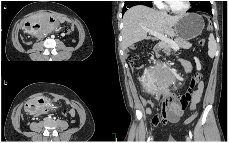
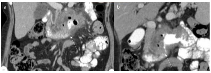
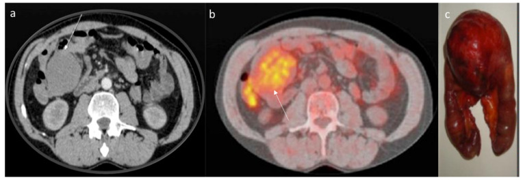
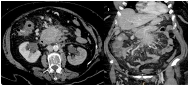
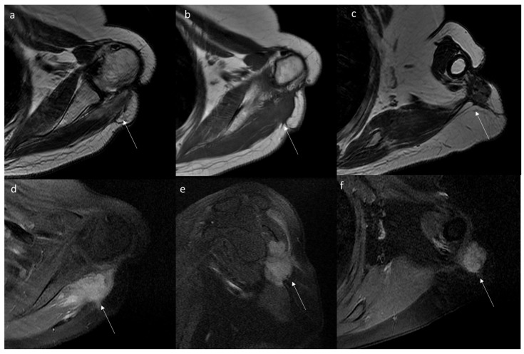
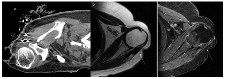
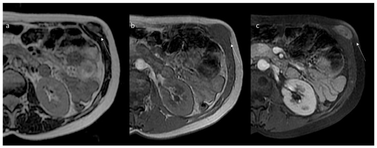
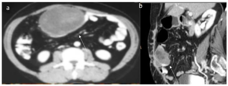
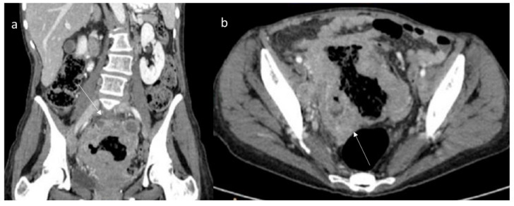
Desmoid tumors (DTs), also known as desmoid fibromatosis or aggressive fibromatosis, are rare, locally invasive, non-metastatic soft tissue tumors. Although histological results represent the gold standard diagnosis, imaging represents the fundamental tool for the diagnosis of these tumors. Although histological analysis represents the gold standard for diagnosis, imaging represents the fundamental tool for the diagnosis of these tumors. DTs represent a challenge for the radiologist, being able to mimic different pathological conditions. A proper diagnosis is required to establish an adequate therapeutic approach. Multimodality imaging, including ultrasound (US), computed tomography (CT) and Magnetic Resonance Imaging (MRI), should be preferred. Different imaging techniques can also guide minimally invasive treatments and monitor their effectiveness. The purpose of this review is to describe the state-of-the-art multidisciplinary imaging of DTs; and its role in patient management.
硬纤维瘤(DTs),也称为韧带样纤维瘤病或侵袭性纤维瘤病,是一种罕见的、局部侵袭性的、非转移性软组织肿瘤。虽然组织学结果是诊断的金标准,但影像学检查是诊断这些肿瘤的基本工具。虽然组织学分析是诊断的金标准,但影像学检查是诊断这些肿瘤的基本工具。硬纤维瘤对放射科医生来说是一个挑战,因为它能够模仿不同的病理状况。需要进行正确的诊断以建立适当的治疗方法。应首选包括超声(US)、计算机断层扫描(CT)和磁共振成像(MRI)在内的多模态成像。不同的成像技术还可以指导微创治疗并监测其有效性。本综述的目的是描述硬纤维瘤的最新多学科成像;及其在患者管理中的作用。