Department of Radiation Oncology, Graduate School of Medicine, Hokkaido University, North-15, West-7, Kita-Ku, Sapporo, Hokkaido, 060-8638, Japan.
Department of Radiation Medical Science and Engineering, Faculty of Medicine, Hokkaido University, North-15, West-7, Kita-Ku, Sapporo, Hokkaido, 060-8638, Japan.
Radiat Oncol. 2019 Dec 12;14(1):226. doi: 10.1186/s13014-019-1424-8.
To determine the best method to contour the planning organ at risk volume (PRV) for the urethra, this study aimed to investigate the displacement of a Foley catheter in the urethra with a soft and thin guide-wire.
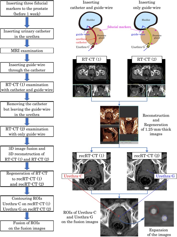
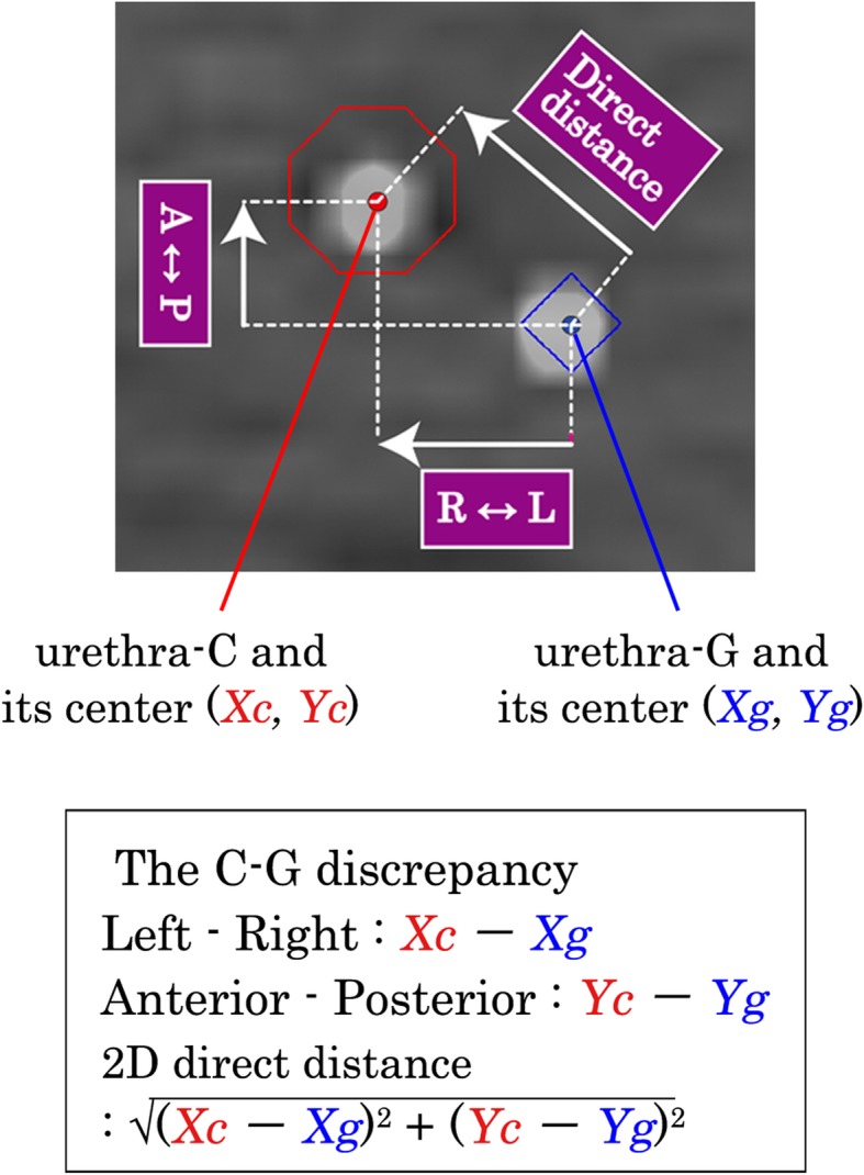
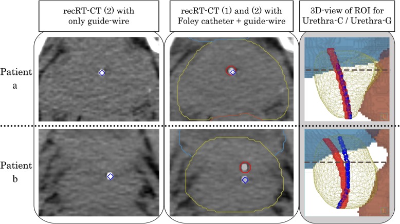
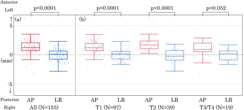
For each patient, the study used two sets of computed tomography (CT) images for radiation treatment planning (RT-CT): (1) set with a Foley urethral catheter (4.0 mm diameter) plus a guide-wire (0.46 mm diameter) in the first RT-CT and (2) set with a guide-wire alone in the second CT recorded 2 min after the first RT-CT. Using three fiducial markers in the prostate for image fusion, the displacement between the catheter and the guide-wire in the prostatic urethra was calculated. In 155 consecutive patients treated between 2011 and 2017, 5531 slices of RT-CT were evaluated.
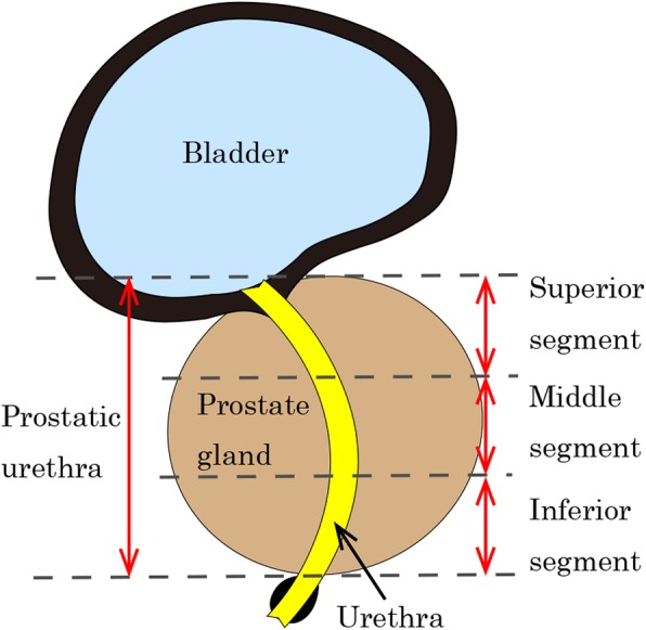
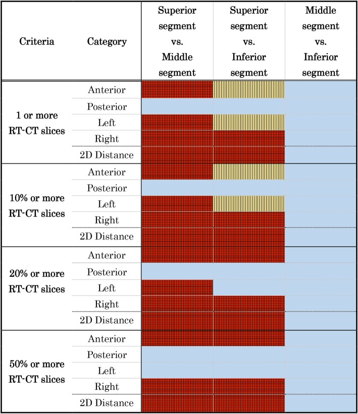
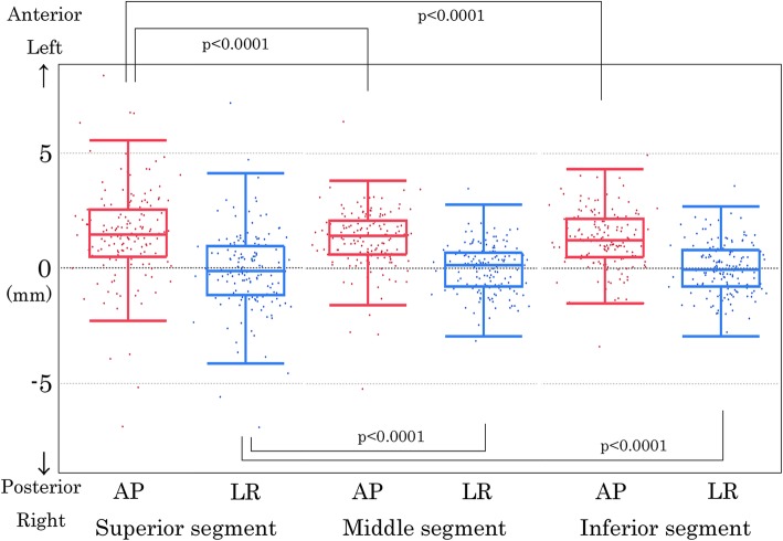
Assuming that ≥3.0 mm of difference between the catheter and the guide-wire position was a significant displacement, the urethra with the catheter was displaced significantly from the urethra with the guide-wire alone in > 20% of the RT-CT slices in 23.2% (36/155) of the patients. The number of patients who showed ≥3.0 mm anterior displacement with the catheter in ≥20% RT-CT slices was significantly larger at the superior segment (38/155) than at the middle (14/155) and inferior segments (18/155) of the prostatic urethra (p < 0.0167).
The urethral position with a Foley catheter is different from the urethral position with a thin and soft guide-wire in a significant proportion of the patients. This should be taken into account for the PRV of the urethra to ensure precise radiotherapy such as in urethra-sparing radiotherapy.
为了确定勾画计划器官受照体积(PRV)中尿道的最佳方法,本研究旨在通过使用柔软细导丝来研究尿道中 Foley 导管的移位。
对于每个患者,研究使用两组放射治疗计划的计算机断层扫描(CT)图像:(1)在第一次 RT-CT 中带有 Foley 尿道导管(4.0mm 直径)和导丝(0.46mm 直径)的一组,(2)在第一次 RT-CT 后 2 分钟单独记录导丝的第二组 CT。通过在前列腺中使用三个基准标记进行图像融合,计算导管和导丝在前列腺尿道中的移位。在 2011 年至 2017 年间治疗的 155 例连续患者中,评估了 5531 张 RT-CT 切片。
假设导管和导丝位置之间的差异≥3.0mm 为显著移位,则在 23.2%(36/155)的患者中,超过 20%的 RT-CT 切片中导管和导丝的尿道位置明显不同。在具有导管的患者中,在前尿道位置≥3.0mm 的患者中,在前尿道上段(38/155)明显多于中尿道(14/155)和下段(18/155)(p<0.0167)。
在相当一部分患者中,带有 Foley 导管的尿道位置与带有柔软细导丝的尿道位置不同。这在为确保精确放疗(如尿道保留放疗)而勾画尿道 PRV 时应予以考虑。