• 文献检索
  • 文档翻译
  • 深度研究
  • 学术资讯
  • Suppr Zotero 插件Zotero 插件
  • 邀请有礼
  • 套餐&价格
  • 历史记录
应用&插件
Suppr Zotero 插件Zotero 插件浏览器插件Mac 客户端Windows 客户端微信小程序
定价
高级版会员购买积分包购买API积分包
服务
文献检索文档翻译深度研究API 文档MCP 服务
关于我们
关于 Suppr公司介绍联系我们用户协议隐私条款
关注我们

Suppr 超能文献

核心技术专利:CN118964589B侵权必究
粤ICP备2023148730 号-1Suppr @ 2026

文献检索

告别复杂PubMed语法,用中文像聊天一样搜索,搜遍4000万医学文献。AI智能推荐,让科研检索更轻松。

立即免费搜索

文件翻译

保留排版,准确专业,支持PDF/Word/PPT等文件格式,支持 12+语言互译。

免费翻译文档

深度研究

AI帮你快速写综述,25分钟生成高质量综述,智能提取关键信息,辅助科研写作。

立即免费体验

使用数字全息显微镜进行定量相位成像可可靠地评估纤维化肠道组织的形态并反映其弹性特性。

Quantitative Phase Imaging Using Digital Holographic Microscopy Reliably Assesses Morphology and Reflects Elastic Properties of Fibrotic Intestinal Tissue.

机构信息

Department of Medicine B for Gastroenterology and Hepatology, University Hospital Muenster, Muenster, Germany.

Institute of Palliative Care, University Hospital Muenster, Muenster, Germany.

出版信息

Sci Rep. 2019 Dec 18;9(1):19388. doi: 10.1038/s41598-019-56045-2.

DOI:10.1038/s41598-019-56045-2
PMID:31852983
原文链接:https://pmc.ncbi.nlm.nih.gov/articles/PMC6920451/
Abstract

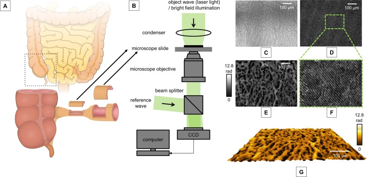
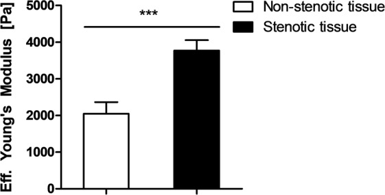
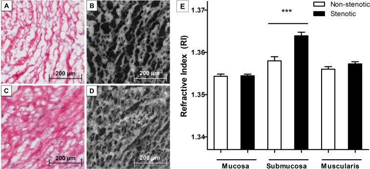
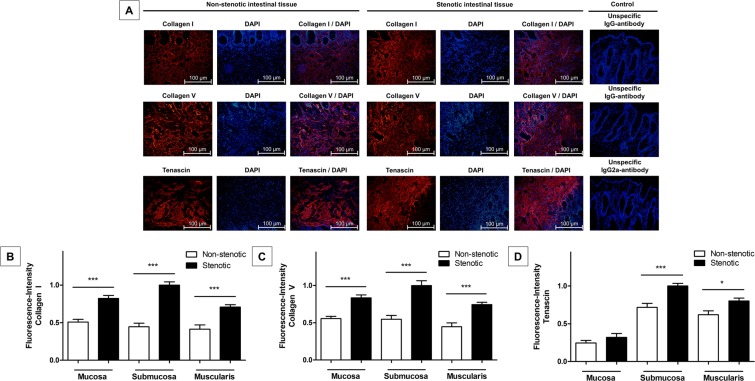
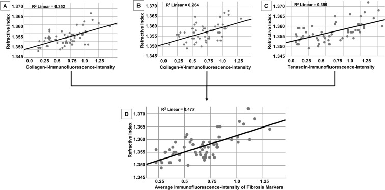
Intestinal strictures are a frequent complication in patients with Crohn's Disease (CD) and the presence of fibrosis within strictures impacts the therapeutic treatment approach. Here, we evaluate quantitative phase imaging (QPI) using digital holographic microscopy (DHM) for the evaluation of fibrosis within CD strictures. 30 full thickness resection specimens were obtained from non-stenotic and stenotic tissue areas of 15 CD patients. Cryostat sections were analyzed by DHM to measure the spatial distribution of the refractive index (RI) to quantify tissue density. Complementary, histopathological evaluation of H&E staining and immunofluorescence (IF) targeting fibrosis markers served as the gold standard. Moreover, tissue stiffness was evaluated by elastography. RI values assessed by DHM were significantly higher in stenotic compared to non-stenotic tissue areas (p < 0.001). Histopathological analysis using H&E staining and IF confirmed the elevated expression of fibrosis markers in stenotic compared to non-stenotic tissue (all p < 0.001). The RI retrieved by DHM strongly correlated with the amount of fibrosis as determined by IF (p < 0.001; R = 0.48). Furthermore, elastography detected a significantly higher tissue stiffness in stenotic as compared to non-stenotic tissue sections (p < 0.001). In conclusion, QPI using DHM accurately assesses fibrotic properties of CD-associated strictures and may improve the characterization of CD strictures.

摘要

肠狭窄是克罗恩病(CD)患者的常见并发症,狭窄处的纤维化存在会影响治疗方法。在这里,我们评估了数字全息显微镜(DHM)定量相位成像(QPI)在 CD 狭窄处纤维化评估中的应用。从 15 例 CD 患者的非狭窄和狭窄组织区域获得 30 个全厚切除标本。通过 DHM 分析冷冻切片,以测量折射率(RI)的空间分布,从而定量组织密度。此外,对 H&E 染色和针对纤维化标志物的免疫荧光(IF)的组织学评估作为金标准。而且,通过弹性成像评估组织硬度。DHM 评估的 RI 值在狭窄组织区域明显高于非狭窄组织区域(p<0.001)。H&E 染色和 IF 组织学分析证实,与非狭窄组织相比,狭窄组织中纤维化标志物的表达升高(均 p<0.001)。DHM 获得的 RI 与 IF 确定的纤维化程度呈强相关性(p<0.001;R=0.48)。此外,弹性成像检测到狭窄组织的组织硬度明显高于非狭窄组织(p<0.001)。总之,DHM 定量相位成像可准确评估 CD 相关狭窄处的纤维化特性,并可能改善 CD 狭窄处的特征描述。

https://cdn.ncbi.nlm.nih.gov/pmc/blobs/8819/6920451/88725ef58f52/41598_2019_56045_Fig5_HTML.jpg
https://cdn.ncbi.nlm.nih.gov/pmc/blobs/8819/6920451/67b5e04e0638/41598_2019_56045_Fig1_HTML.jpg
https://cdn.ncbi.nlm.nih.gov/pmc/blobs/8819/6920451/a19d018ad679/41598_2019_56045_Fig2_HTML.jpg
https://cdn.ncbi.nlm.nih.gov/pmc/blobs/8819/6920451/048bfeeab444/41598_2019_56045_Fig3_HTML.jpg
https://cdn.ncbi.nlm.nih.gov/pmc/blobs/8819/6920451/5cb5cf94a83e/41598_2019_56045_Fig4_HTML.jpg
https://cdn.ncbi.nlm.nih.gov/pmc/blobs/8819/6920451/88725ef58f52/41598_2019_56045_Fig5_HTML.jpg

相似文献

1
Quantitative Phase Imaging Using Digital Holographic Microscopy Reliably Assesses Morphology and Reflects Elastic Properties of Fibrotic Intestinal Tissue.使用数字全息显微镜进行定量相位成像可可靠地评估纤维化肠道组织的形态并反映其弹性特性。
Sci Rep. 2019 Dec 18;9(1):19388. doi: 10.1038/s41598-019-56045-2.
2
Real-time elastography for the detection of fibrotic and inflammatory tissue in patients with stricturing Crohn's disease.实时弹性成像技术在狭窄性克罗恩病患者中检测纤维化和炎症组织的应用
J Ultrasound. 2017 Oct 26;20(4):273-284. doi: 10.1007/s40477-017-0265-2. eCollection 2017 Dec.
3
Real-Time Shear Wave Ultrasound Elastography Differentiates Fibrotic from Inflammatory Strictures in Patients with Crohn's Disease.实时剪切波超声弹性成像可区分克罗恩病患者的纤维化与炎症性狭窄。
Inflamm Bowel Dis. 2018 Sep 15;24(10):2183-2190. doi: 10.1093/ibd/izy115.
4
Bowel stiffness associated with histopathologic scoring of stenosis in patients with Crohn's disease.与克罗恩病患者狭窄的组织病理学评分相关的肠僵硬。
Acta Biomater. 2021 Aug;130:332-342. doi: 10.1016/j.actbio.2021.06.011. Epub 2021 Jun 10.
5
Multimodal Quantitative Phase Imaging with Digital Holographic Microscopy Accurately Assesses Intestinal Inflammation and Epithelial Wound Healing.利用数字全息显微镜进行的多模态定量相位成像可准确评估肠道炎症和上皮伤口愈合。
J Vis Exp. 2016 Sep 13(115):54460. doi: 10.3791/54460.
6
Strain assessment in surgically resected inflammatory and neoplastic bowel lesions.手术切除的炎症性和肿瘤性肠病变的应变评估。
Ultraschall Med. 2014 Apr;35(2):149-58. doi: 10.1055/s-0032-1325535. Epub 2012 Nov 15.
7
Bowel Thickening in Crohn's Disease: Fibrosis or Inflammation? Diagnostic Ultrasound Imaging Tools.克罗恩病中的肠壁增厚:纤维化还是炎症?诊断性超声成像工具
Inflamm Bowel Dis. 2017 Jan;23(1):23-34. doi: 10.1097/MIB.0000000000000997.
8
Noninvasive Multimodal Methods to Differentiate Inflamed vs Fibrotic Strictures in Patients With Crohn's Disease.采用非侵入性多模态方法对克罗恩病患者的炎症性与纤维化性狭窄进行鉴别。
Clin Gastroenterol Hepatol. 2019 Nov;17(12):2397-2415. doi: 10.1016/j.cgh.2019.04.025. Epub 2019 Apr 14.
9
Application of Ultrasound Elastography for Assessing Intestinal Fibrosis in Inflammatory Bowel Disease: Fiction or Reality?超声弹性成像技术在炎症性肠病肠纤维化评估中的应用:是幻想还是现实?
Curr Drug Targets. 2021;22(3):347-355. doi: 10.2174/1389450121666201119142919.
10
Inhibition of Fibroblast Activation Protein Restores a Balanced Extracellular Matrix and Reduces Fibrosis in Crohn's Disease Strictures Ex Vivo.体外抑制成纤维细胞激活蛋白可恢复平衡的细胞外基质并减少克罗恩病狭窄处的纤维化。
Inflamm Bowel Dis. 2018 Jan 18;24(2):332-345. doi: 10.1093/ibd/izx008.

引用本文的文献

1
Measurement of HbA1c with a new phase function using RBC phase image from a low-cost digital holographic microscopy.使用来自低成本数字全息显微镜的红细胞相位图像,通过一种新的相位函数测量糖化血红蛋白(HbA1c)。
Biotechnol Lett. 2025 Jun 23;47(4):65. doi: 10.1007/s10529-025-03610-7.
2
Revealing 3D microanatomical structures of unlabeled thick cancer tissues using holotomography and virtual H&E staining.利用全层析成像和虚拟苏木精-伊红染色揭示未标记厚癌组织的三维微观解剖结构。
Nat Commun. 2025 May 22;16(1):4781. doi: 10.1038/s41467-025-59820-0.
3
White Light Diffraction Phase Microscopy in Imaging of Breast and Colon Tissues.

本文引用的文献

1
Histopathology Scoring Systems of Stenosis Associated With Small Bowel Crohn's Disease: A Systematic Review.狭窄相关的小肠克罗恩病的组织病理学评分系统:系统评价。
Gastroenterology. 2020 Jan;158(1):137-150.e1. doi: 10.1053/j.gastro.2019.08.033. Epub 2019 Aug 30.
2
Assessment of Crohn's disease-associated small bowel strictures and fibrosis on cross-sectional imaging: a systematic review.基于横断面成像的克罗恩病相关小肠狭窄和纤维化评估:系统综述。
Gut. 2019 Jun;68(6):1115-1126. doi: 10.1136/gutjnl-2018-318081. Epub 2019 Apr 3.
3
Nano-optic endoscope for high-resolution optical coherence tomography .
白光衍射相显微镜在乳腺和结肠组织成像中的应用
Diagnostics (Basel). 2024 Sep 6;14(17):1966. doi: 10.3390/diagnostics14171966.
4
Grading of glioma tumors using digital holographic microscopy.使用数字全息显微镜对胶质瘤肿瘤进行分级。
Heliyon. 2024 Apr 23;10(9):e29897. doi: 10.1016/j.heliyon.2024.e29897. eCollection 2024 May 15.
5
Recent Advances and Current Trends in Transmission Tomographic Diffraction Microscopy.透射断层衍射显微镜的最新进展与当前趋势
Sensors (Basel). 2024 Feb 29;24(5):1594. doi: 10.3390/s24051594.
6
Microindentation of fresh soft biological tissue: A rapid tissue sectioning and mounting protocol.新鲜软生物组织的微压痕:一种快速的组织切片和安装方案。
PLoS One. 2024 Feb 29;19(2):e0297618. doi: 10.1371/journal.pone.0297618. eCollection 2024.
7
Artificial intelligence-enabled quantitative phase imaging methods for life sciences.人工智能赋能的生命科学定量相位成像方法。
Nat Methods. 2023 Nov;20(11):1645-1660. doi: 10.1038/s41592-023-02041-4. Epub 2023 Oct 23.
8
Quantitative Phase Imaging Using Digital Holographic Microscopy to Assess the Degree of Intestinal Inflammation in Patients with Ulcerative Colitis.使用数字全息显微镜进行定量相位成像以评估溃疡性结肠炎患者的肠道炎症程度
J Clin Med. 2023 Jun 15;12(12):4067. doi: 10.3390/jcm12124067.
9
Quantification of structural heterogeneity in H&E stained clear cell renal cell carcinoma using refractive index tomography.使用折射率断层扫描对苏木精-伊红染色的透明细胞肾细胞癌中的结构异质性进行定量分析。
Biomed Opt Express. 2023 Feb 7;14(3):1071-1081. doi: 10.1364/BOE.484092. eCollection 2023 Mar 1.
10
Long-term label-free assessments of individual bacteria using three-dimensional quantitative phase imaging and hydrogel-based immobilization.使用三维定量相衬成像和水凝胶固定化技术对单个细菌进行长期无标记评估。
Sci Rep. 2023 Jan 2;13(1):46. doi: 10.1038/s41598-022-27158-y.
用于高分辨率光学相干断层扫描的纳米光学内窥镜
Nat Photonics. 2018 Sep;12(9):540-547. doi: 10.1038/s41566-018-0224-2. Epub 2018 Jul 30.
4
Efficacy of Endoscopic Dilation of Gastroduodenal Crohn's Disease Strictures: A Systematic Review and Meta-Analysis of Individual Patient Data.内镜下扩张治疗胃十二指肠克罗恩病狭窄的疗效:一项个体患者数据的系统评价和荟萃分析。
Clin Gastroenterol Hepatol. 2019 Nov;17(12):2514-2522.e8. doi: 10.1016/j.cgh.2018.11.048. Epub 2018 Nov 29.
5
An expert consensus to standardise definitions, diagnosis and treatment targets for anti-fibrotic stricture therapies in Crohn's disease.抗纤维化狭窄治疗克罗恩病的专家共识:定义、诊断和治疗目标的标准化。
Aliment Pharmacol Ther. 2018 Aug;48(3):347-357. doi: 10.1111/apt.14853. Epub 2018 Jun 19.
6
Crohn's disease associated strictures.克罗恩病相关狭窄。
J Gastroenterol Hepatol. 2018 May;33(5):998-1008. doi: 10.1111/jgh.14119. Epub 2018 Mar 12.
7
Consensus Recommendations for Evaluation, Interpretation, and Utilization of Computed Tomography and Magnetic Resonance Enterography in Patients With Small Bowel Crohn's Disease.《小肠克罗恩病患者计算机断层扫描和磁共振肠造影评估、解读和应用的共识建议》。
Radiology. 2018 Mar;286(3):776-799. doi: 10.1148/radiol.2018171737. Epub 2018 Jan 10.
8
Quantitative phase microscopy for evaluation of intestinal inflammation and wound healing utilizing label-free biophysical markers.利用无标记生物物理标志物的定量相显微镜评估肠道炎症和伤口愈合
Histol Histopathol. 2018 May;33(5):417-432. doi: 10.14670/HH-11-937. Epub 2017 Oct 9.
9
Validity of Contrast-enhanced Ultrasonography and Dynamic Contrast-enhanced MR Enterography in the Assessment of Transmural Activity and Fibrosis in Crohn's Disease.对比增强超声和动态对比增强磁共振肠道成像在评估克罗恩病肠壁活动度和纤维化中的有效性。
J Crohns Colitis. 2018 Jan 5;12(1):48-56. doi: 10.1093/ecco-jcc/jjx111.
10
Endoscopic optical coherence tomography: technologies and clinical applications [Invited].内镜光学相干断层扫描:技术与临床应用[特邀文章]
Biomed Opt Express. 2017 Apr 7;8(5):2405-2444. doi: 10.1364/BOE.8.002405. eCollection 2017 May 1.