Ito Taeko, Inui Hiroshi, Miyasaka Toshiteru, Shiozaki Tomoyuki, Hasukawa Akihito, Yamanaka Toshiaki, Kichikawa Kimihiko, Kitahara Tadashi
Department of Otolaryngology-Head and Neck Surgery Nara Medical University Kashihara Nara Japan.
Inui ENT Clinic Sakurai Nara Japan.
Laryngoscope Investig Otolaryngol. 2019 Nov 7;4(6):653-658. doi: 10.1002/lio2.313. eCollection 2019 Dec.
Recently, 3-Tesla magnetic resonance imaging (MRI) with intravenous gadolinium injection has been used to reveal endolymphatic hydrops (EH). In the present study, we aimed to evaluate EH in patients with Meniere's disease (MD) objectively and quantitatively, and compared the endolymphatic space (ELS) in individuals with MD and healthy controls, to gain understanding of the characteristics of MD.
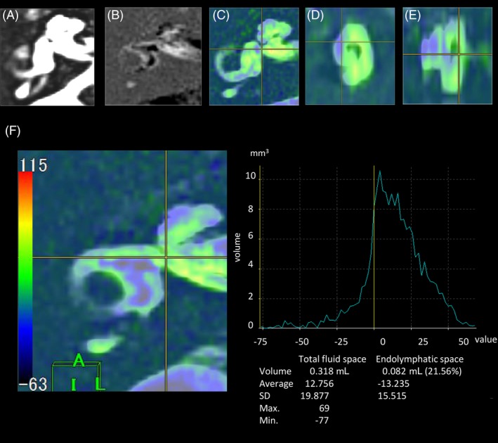
Eighty-two patients with unilateral MD (uMD), 16 patients with bilateral MD (bMD), and 47 healthy volunteers were enrolled. All participants underwent 3-T MRI at 4 hours after intravenous gadolinium injection. The volumes of the total fluid space (TFS) and ELS were measured semiautomatically using our workstation, and the percentage of ELS to TFS (ELS percentage) was calculated.
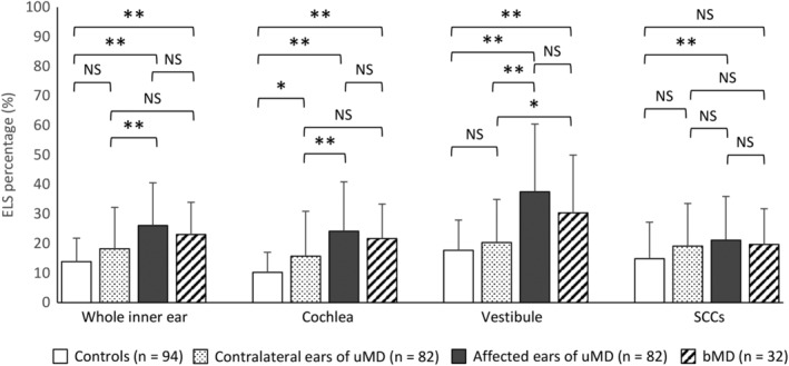
The ELS percentage was 13.9 in the ears of controls, 18.2 in the contralateral ear of individuals with uMD, 26.1 in the affected ears of these individuals, and 23.0 in both ears of individuals with bMD. The ELS percentages in the affected ear of uMD and the ears of bMD individuals were significantly higher than that in the ears of control individuals ( < .01, one-way analysis of variance (ANOVA), Tukey's test).
The ELS is significantly larger in the affected ears of uMD and in both ears of bMD individuals. Accurate diagnosis of MD can be facilitated by using 3-T MRI 4 hours after intravenous gadolinium injection and performing volumetric measurements of the ELS.
2b.
最近,静脉注射钆对比剂的3特斯拉磁共振成像(MRI)已被用于揭示内淋巴积水(EH)。在本研究中,我们旨在客观、定量地评估梅尼埃病(MD)患者的EH情况,并比较MD患者和健康对照者的内淋巴间隙(ELS),以了解MD的特征。
纳入82例单侧MD(uMD)患者、16例双侧MD(bMD)患者和47名健康志愿者。所有参与者在静脉注射钆对比剂4小时后接受3-T MRI检查。使用我们的工作站半自动测量总液体间隙(TFS)和ELS的体积,并计算ELS占TFS的百分比(ELS百分比)。
对照组耳朵的ELS百分比为13.9,uMD患者对侧耳朵的ELS百分比为18.2,这些患者患侧耳朵的ELS百分比为26.1,bMD患者双耳的ELS百分比为23.0。uMD患者患侧耳朵和bMD患者耳朵的ELS百分比显著高于对照组耳朵(<0.01,单因素方差分析(ANOVA),Tukey检验)。
uMD患者患侧耳朵和bMD患者双耳的ELS明显更大。静脉注射钆对比剂4小时后使用3-T MRI并对ELS进行容积测量有助于MD的准确诊断。
2b。