Yektaeian Narjes, Mehrabani Davood, Sepaskhah Mozhdeh, Zare Shahrokh, Jamhiri Iman, Hatam Gholamreza
Department of Parasitology and Mycology, School of Medicine, Shiraz University of Medical Sciences, Shiraz, Iran.
Stem Cells Technology Research Center, Shiraz University of Medical Sciences, Shiraz, Iran.
Heliyon. 2019 Dec 18;5(12):e03073. doi: 10.1016/j.heliyon.2019.e03073. eCollection 2019 Dec.
This study aims to evaluate the use of fluorescent dye Dil and super vital dye acridine orange (AO) tracking of labeled in the fibroblast cells.
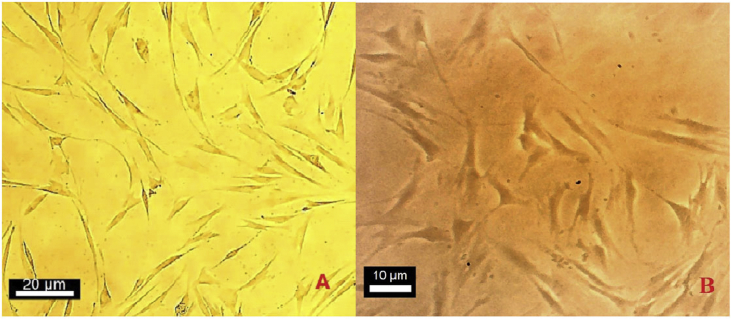
Dil crystal and AO were used to stain in a co-culture of the fibroblasts with the parasite. AO staining solution was added to 1 × 10 parasites. After 10 min, the stained parasites were centrifuged and washed seven times with phosphate buffered saline (PBS). The stained promastigote was incubated with fibroblasts for 6-8 h. The presence of stained parasites with AO in the fibroblast was assessed using a fluorescence microscope. 1 × 10/mL promastigote of was gently suspended and mixed by Dil staining solution with an ultimate concentration of 0.002 μg/mL and it was kept for 20 min at the room temperature. Subsequently, after washing it in PBS for seven times, it was centrifuged at 3000 rpm for 10 min. The supernatant was removed and the precipitate containing stained promastigote was suspended in fresh DMEM F12 with fibroblasts at 37 °C for 6 h. The presence of stained parasites with Dil in fibroblast was assessed using a fluorescence microscope. Fibroblast characterization was undertaken by a real-time polymerase chain reaction (PCR).
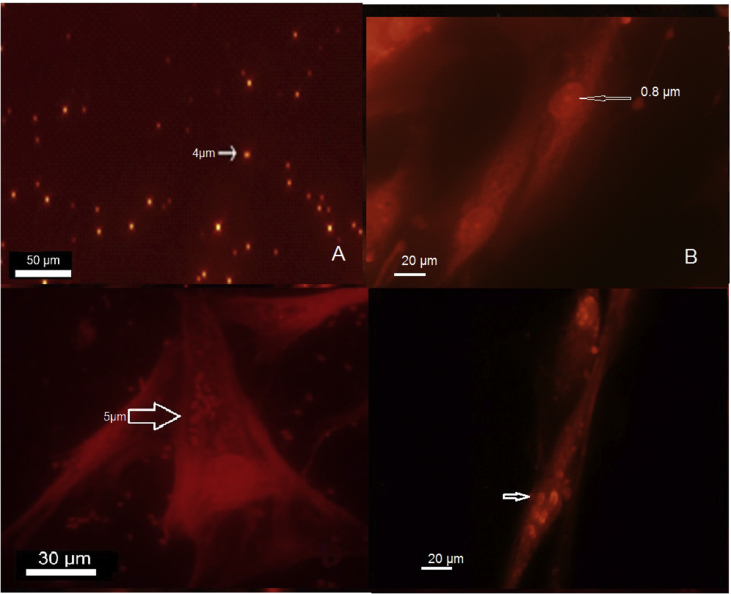
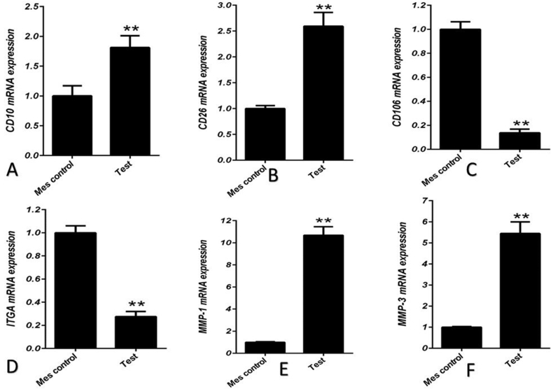
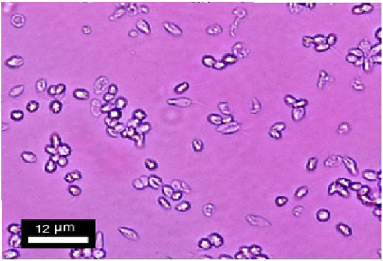
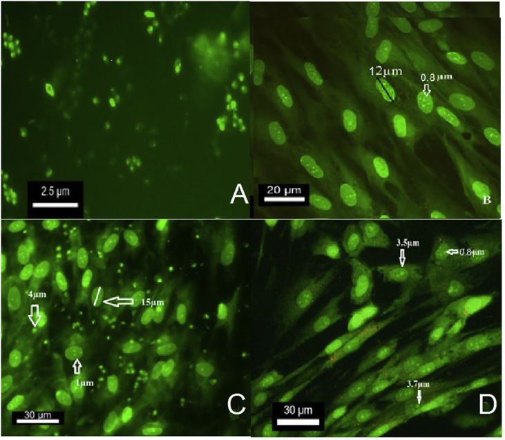
Acridine orange staining assisted in detection of the live parasite in the fibroblast cells. Free promastigote looked green before entering into the fibroblasts after 12 h culture. The parasite entered the cytoplasm of fibroblasts at the beginning of the exposure and gradually entered the nucleus of the fibroblast. The fibroblast nucleus was entirely stained green by AO. The appeared green under the fluorescent microscope. Dil staining revealed that the internalized parasites with red/orange color were localized within the cytoplasm after 6-hours and the nucleus of the fibroblasts after 72-hours following culture. Human fibroblasts were positive at the expression of CD10, CD26, matrix metalloproteinase-1 (MMP-1) and matrix metalloproteinase-3 (MMP-3) and negative for CD106 and integrin alpha 11.
The fluorescent dye Dil staining is a safe, easy to use, inexpensive and fast method for labeling of the parasite in the fibroblast cells. Acridine orange staining could be useful for tracing the parasites in the fibroblasts too. In this study, both Dil and AO were compared and considered as suitable vital dyes for identifying labeled in the fibroblast , but Dil was superior to AO with its feature does not transfer from the labeled to unlabeled cells.
本研究旨在评估使用荧光染料Dil和超活染料吖啶橙(AO)对成纤维细胞中标记的寄生虫进行追踪。
使用Dil晶体和AO对寄生虫与成纤维细胞的共培养物进行染色。将AO染色溶液加入1×10个寄生虫中。10分钟后,对染色后的寄生虫进行离心,并用磷酸盐缓冲盐水(PBS)洗涤7次。将染色后的前鞭毛体与成纤维细胞孵育6 - 8小时。使用荧光显微镜评估成纤维细胞中被AO染色的寄生虫的存在情况。将1×10/mL的前鞭毛体轻轻悬浮,并用终浓度为0.002μg/mL的Dil染色溶液混合,在室温下放置20分钟。随后,用PBS洗涤7次后,以3000 rpm离心10分钟。去除上清液,将含有染色后前鞭毛体的沉淀物悬浮于新鲜的DMEM F12中,与成纤维细胞在37℃下孵育6小时。使用荧光显微镜评估成纤维细胞中被Dil染色的寄生虫的存在情况。通过实时聚合酶链反应(PCR)进行成纤维细胞表征。
吖啶橙染色有助于检测成纤维细胞中的活寄生虫。在培养12小时后,游离的前鞭毛体在进入成纤维细胞之前呈绿色。在接触开始时,寄生虫进入成纤维细胞的细胞质,并逐渐进入成纤维细胞的细胞核。成纤维细胞的细胞核被AO完全染成绿色。在荧光显微镜下呈绿色。Dil染色显示,内化的寄生虫在培养6小时后呈红色/橙色,位于细胞质内,培养72小时后位于成纤维细胞的细胞核内。人成纤维细胞在CD10、CD26、基质金属蛋白酶 - 1(MMP - 1)和基质金属蛋白酶 - 3(MMP - 3)的表达上呈阳性,而在CD106和整合素α11上呈阴性。
荧光染料Dil染色是一种安全、易于使用、廉价且快速的方法,可用于标记成纤维细胞中的寄生虫。吖啶橙染色也可用于追踪成纤维细胞中的寄生虫。在本研究中,对Dil和AO进行了比较,认为它们都是用于识别成纤维细胞中标记寄生虫的合适活染料,但Dil优于AO,其特点是不会从标记细胞转移到未标记细胞。