Department of Pharmacology, Yong Loo Lin School of Medicine, National University of Singapore, Singapore, 119260, Singapore.
Department of Anatomy, Yong Loo Lin School of Medicine, National University of Singapore, Singapore, 119260, Singapore.
Neuromolecular Med. 2020 Jun;22(2):293-303. doi: 10.1007/s12017-019-08586-y. Epub 2020 Jan 4.
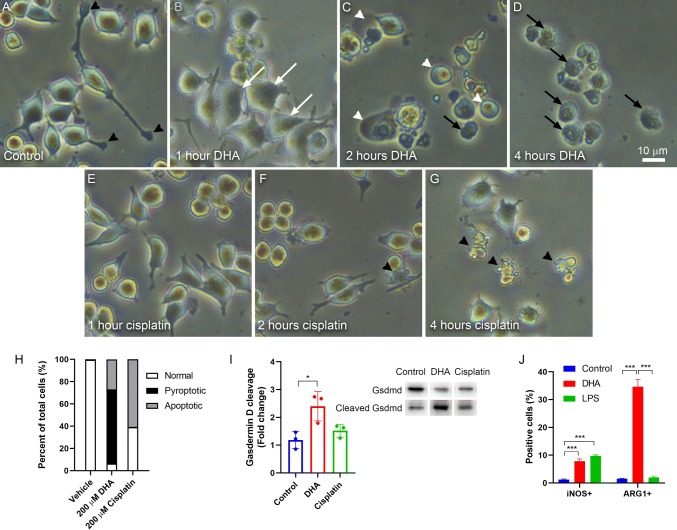
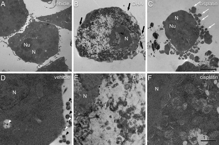
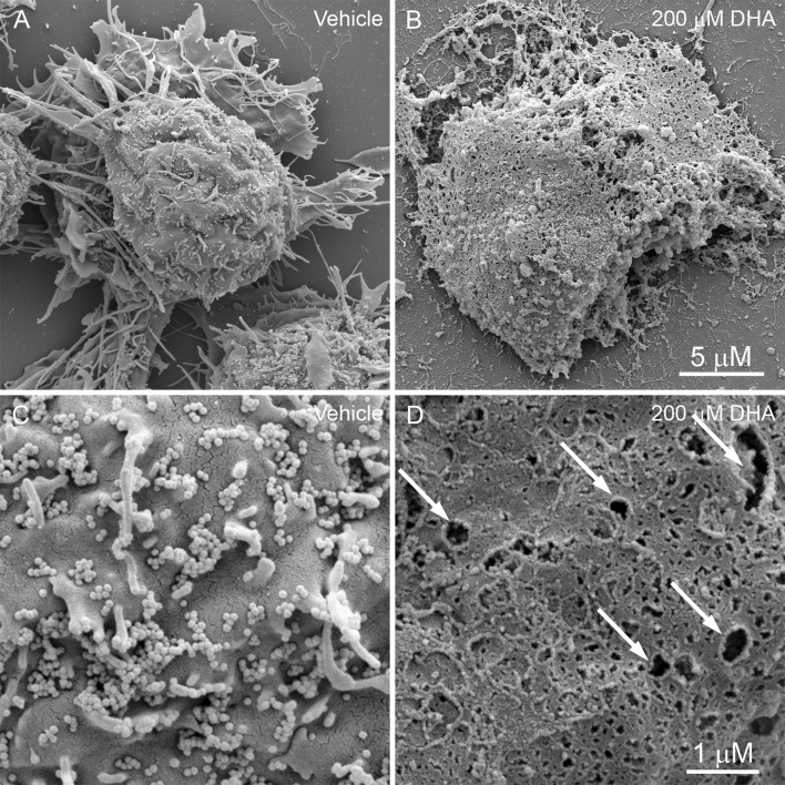
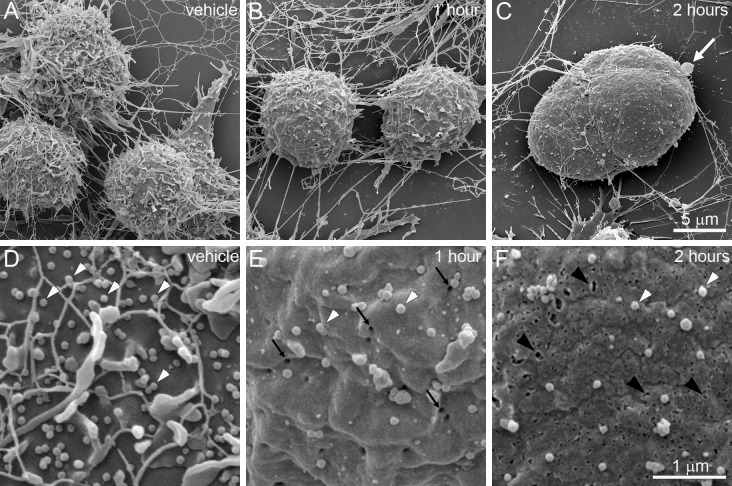
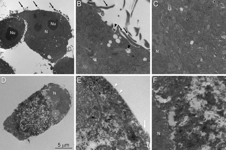
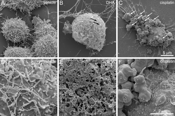
Microglial cells are resident macrophages of the central nervous system (CNS) that respond to bioactive lipids such as docosahexaenoic acid (DHA). Low micromolar concentrations of DHA typically promote anti-inflammatory functions of microglia, but higher concentrations result in a form of pro-inflammatory programmed cell death known as pyroptosis. This study used scanning electron microscopy (SEM) and transmission electron microscopy (TEM) to investigate the morphological characteristics of pyroptosis in BV-2 microglial cells following exposure to 200 µM DHA. Vehicle-treated cells are characterized by extended processes, spine-like projections or 0.4 to 5.2 µm in length, and numerous extracellular vesicles (EVs) tethered to the surface of the plasma membrane. In contrast to vehicle-treated cells, gross abnormalities are observed after treating cells with 200 µM DHA for 4 h. These include the appearance of numerous pits or pores of varying sizes across the cell surface, structural collapse and flattening of the cell shape. Moreover, EVs and spines were lost following DHA treatment, possibly due to release from the cell surface. The membrane pores appear after DHA treatment initially measured ~ 30 nm, consistent with the previously reported gasdermin D (GSDMD) pore complexes. Complete collapse of cytoplasmic organization and loss of nuclear envelope integrity were also observed in DHA-treated cells. These processes are morphologically distinct from the changes that occur during cisplatin-induced apoptosis, such as the appearance of apoptotic bodies and tightly packed organelles, and the maintenance of EVs and nuclear envelope integrity. Cumulatively, this study provides a systematic description of the ultrastructural characteristics of DHA-induced pyroptosis, including distinguishing features that differentiate this process from apoptosis.
小胶质细胞是中枢神经系统 (CNS) 的常驻巨噬细胞,可对生物活性脂质(如二十二碳六烯酸 (DHA))做出反应。低微摩尔浓度的 DHA 通常可促进小胶质细胞的抗炎功能,但较高浓度则会导致一种称为细胞焦亡的促炎程序性细胞死亡形式。本研究使用扫描电子显微镜 (SEM) 和透射电子显微镜 (TEM) 来研究暴露于 200µM DHA 后 BV-2 小胶质细胞中细胞焦亡的形态特征。用载体处理的细胞的特征是具有延伸的突起、棘状突起或长 0.4 至 5.2µm,以及许多连接到质膜表面的细胞外囊泡 (EV)。与用载体处理的细胞相比,用 200µM DHA 处理细胞 4 小时后会观察到明显的异常。这些异常包括在整个细胞表面出现许多大小不一的凹坑或孔、细胞形状的结构坍塌和平坦化。此外,EV 和棘突在 DHA 处理后丢失,可能是由于从细胞表面释放。DHA 处理后出现的膜孔最初测量约为 30nm,与先前报道的 Gasdermin D (GSDMD) 孔复合物一致。在 DHA 处理的细胞中还观察到细胞质组织的完全崩溃和核膜完整性的丧失。这些过程在形态上与顺铂诱导的细胞凋亡过程中发生的变化不同,例如凋亡小体的出现和紧密堆积的细胞器,以及 EV 和核膜完整性的维持。总之,本研究提供了 DHA 诱导的细胞焦亡的超微结构特征的系统描述,包括区分该过程与细胞凋亡的特征。