LGEI, IMT Mines Alès, Institut Mines-Télécom et Université de Montpellier Sud de France, 6 Avenue de Clavières, 30100 Alès, France.
INRA, SPE, 400 route des Chappes BP 167, 06903 Sophia-Antipolis CEDEX, France.
Viruses. 2020 Jan 2;12(1):50. doi: 10.3390/v12010050.
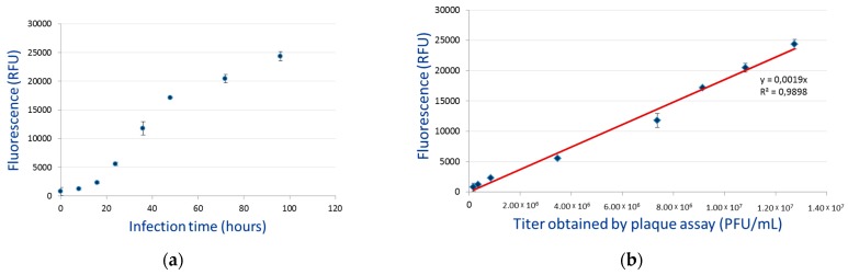
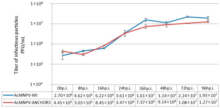
Many steps in the baculovirus life cycle, from initial ingestion to the subsequent infection of all larval cells, remain largely unknown; primarily because it has hitherto not been possible to follow individual genomes and their lineages. Use of ANCHOR technology allows a high intensity fluorescent labelling of DNA. When applied to a virus genome, it is possible to follow individual particles, and the overall course of infection. This technology has been adapted to enable labelling of the baculovirus Autographa californica Multiple NucleoPolyhedroVirus genome, as a first step to its application to other baculoviruses. AcMNPV was modified by inserting the two components of ANCHOR: a specific DNA-binding protein fused to a fluorescent reporter, and the corresponding DNA recognition sequence. The resulting modified virus was stable, infectious, and replicated correctly in 9 (Sf9) cells and in vivo. Both budded viruses and occlusion bodies were clearly distinguishable, and infecting cells or larvae allowed the infection process to be monitored in living cells or tissues. The level of fluorescence in the culture medium of infected cells in vitro showed a good correlation with the number of infectious budded viruses. A cassette that can be used in other baculoviruses has been designed. Altogether our results introduce for the first time the generation of autofluorescent baculovirus and their application to follow infection dynamics directly in living cells or tissues.
杆状病毒生活史的许多步骤,从最初的摄入到随后感染所有幼虫细胞,仍然很大程度上未知;主要是因为迄今为止不可能跟踪单个基因组及其谱系。使用 ANCHOR 技术可以对 DNA 进行高强度荧光标记。当应用于病毒基因组时,可以跟踪单个颗粒及其整体感染过程。该技术已被改编为标记杆状病毒 Autographa californica 多核多角体病毒基因组,作为将其应用于其他杆状病毒的第一步。通过插入 ANCHOR 的两个组件:融合到荧光报告蛋白的特定 DNA 结合蛋白和相应的 DNA 识别序列,对 AcMNPV 进行了修饰。由此产生的修饰病毒稳定、具有感染力,并在 Sf9 细胞和体内正确复制。出芽病毒和包埋体都可以清晰地区分,感染细胞或幼虫可以在活细胞或组织中监测感染过程。体外感染细胞培养基中的荧光强度与感染性出芽病毒的数量之间存在良好的相关性。已经设计了一个可用于其他杆状病毒的盒。总的来说,我们的结果首次引入了自发荧光杆状病毒的产生,并将其直接应用于活细胞或组织中感染动力学的跟踪。