The Laboratory for Therapeutic 3D Bioprinting, Department of Orthopaedic Surgery, Massachusetts General Hospital, Harvard Medical School, Boston, MA 02114, USA.
Int J Mol Sci. 2020 Jan 2;21(1):315. doi: 10.3390/ijms21010315.
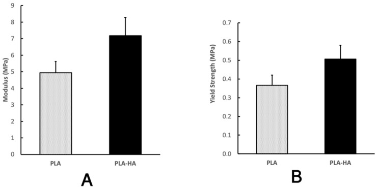
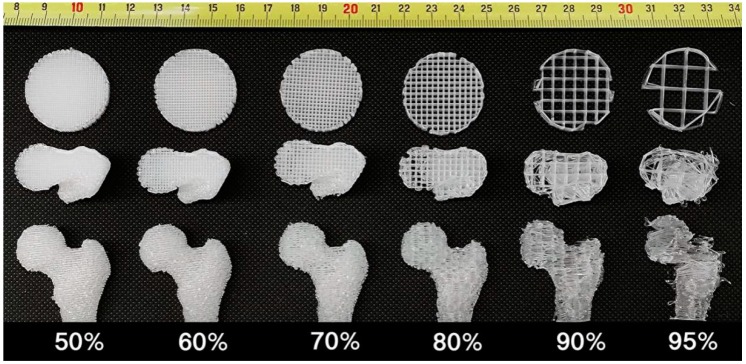
Fused deposit modeling (FDM) 3D printing technology cannot generate scaffolds with high porosity while maintaining good integrity, anatomical-surface detail, or high surface area-to-volume ratio (S/V). Solvent casting and particulate leaching (SCPL) technique generates scaffolds with high porosity and high S/V. However, it is challenging to generate complex-shaped scaffolds; and solvent, particle and residual water removal are time consuming. Here we report techniques surmounting these problems, successfully generating a highly porous scaffold with the anatomical-shape characteristics of a human femur by polylactic acid polymer (PLA) and PLA-hydroxyapatite (HA) casting and salt leaching. The mold is water soluble and is easily removable. By perfusing with ethanol, water, and dry air sequentially, the solvent, salt, and residual water were removed 20 fold faster than utilizing conventional methods. The porosities are uniform throughout the femoral shaped scaffold generated with PLA or PLA-HA. Both scaffolds demonstrated good biocompatibility with the pre-osteoblasts (MC3T3-E1) fully attaching to the scaffold within 8 h. The cells demonstrated high viability and proliferation throughout the entire time course. The HA-incorporated scaffolds demonstrated significantly higher compressive strength, modulus and osteoinductivity as evidenced by higher levels of alkaline-phosphatase activity and calcium deposition. When 3D printing a 3D model at 95% porosity or above, our technology preserves integrity and surface detail when compared with FDM-generated scaffolds. Our technology can also generate scaffolds with a 31 fold larger S/V than FDM. We have developed a technology that is a versatile tool in creating personalized, patient-specific bone graft scaffolds efficiently with high porosity, good scaffold integrity, high anatomical-shaped surface detail and large S/V.
熔融沉积成型(FDM)3D 打印技术无法在保持完整性的同时生成具有高孔隙率的支架,也无法兼顾解剖表面细节或高表面积体积比(S/V)。溶剂浇铸和颗粒沥滤(SCPL)技术可生成具有高孔隙率和高 S/V 的支架。但是,制造复杂形状的支架具有挑战性,并且去除溶剂、颗粒和残留水需要时间。在这里,我们报告了克服这些问题的技术,成功地通过聚乳酸聚合物(PLA)和 PLA-羟基磷灰石(HA)浇铸和盐沥滤生成具有人体股骨解剖形状特征的高多孔支架。模具是水溶性的,易于去除。通过依次灌注乙醇、水和干燥空气,溶剂、盐和残留水的去除速度比传统方法快 20 倍。PLA 或 PLA-HA 生成的股骨形状支架的孔隙率在整个支架中均匀。两种支架均表现出良好的生物相容性,前成骨细胞(MC3T3-E1)在 8 小时内完全附着在支架上。细胞在整个时间过程中均表现出高活力和增殖。HA 掺入的支架表现出更高的抗压强度、模量和骨诱导性,这表现为碱性磷酸酶活性和钙沉积水平更高。当以 95%或更高的孔隙率 3D 打印 3D 模型时,与 FDM 生成的支架相比,我们的技术可保持完整性和表面细节。与 FDM 相比,我们的技术还可以生成 S/V 大 31 倍的支架。我们已经开发出一种技术,该技术是一种通用工具,可以有效地制造具有高孔隙率、良好支架完整性、高解剖形状表面细节和大 S/V 的个性化、患者特异性骨移植物支架。