Department of Biology, Georgia State University, Atlanta, GA, USA.
Department of Computer Science, Georgia State University, Atlanta, GA, USA.
FEBS J. 2020 Aug;287(15):3235-3254. doi: 10.1111/febs.15207. Epub 2020 Jan 23.
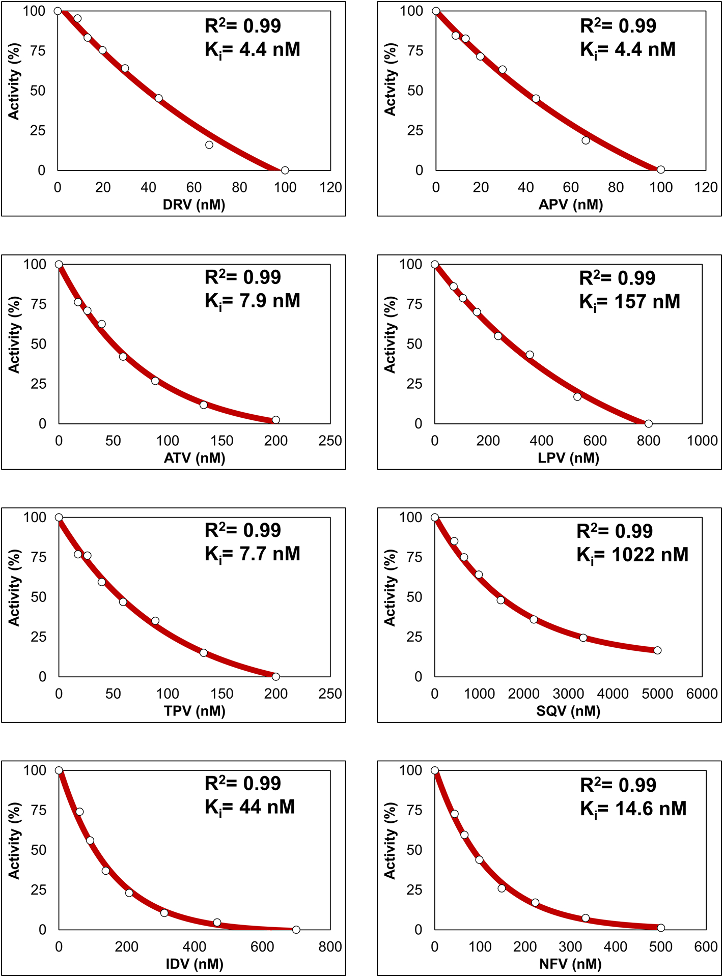
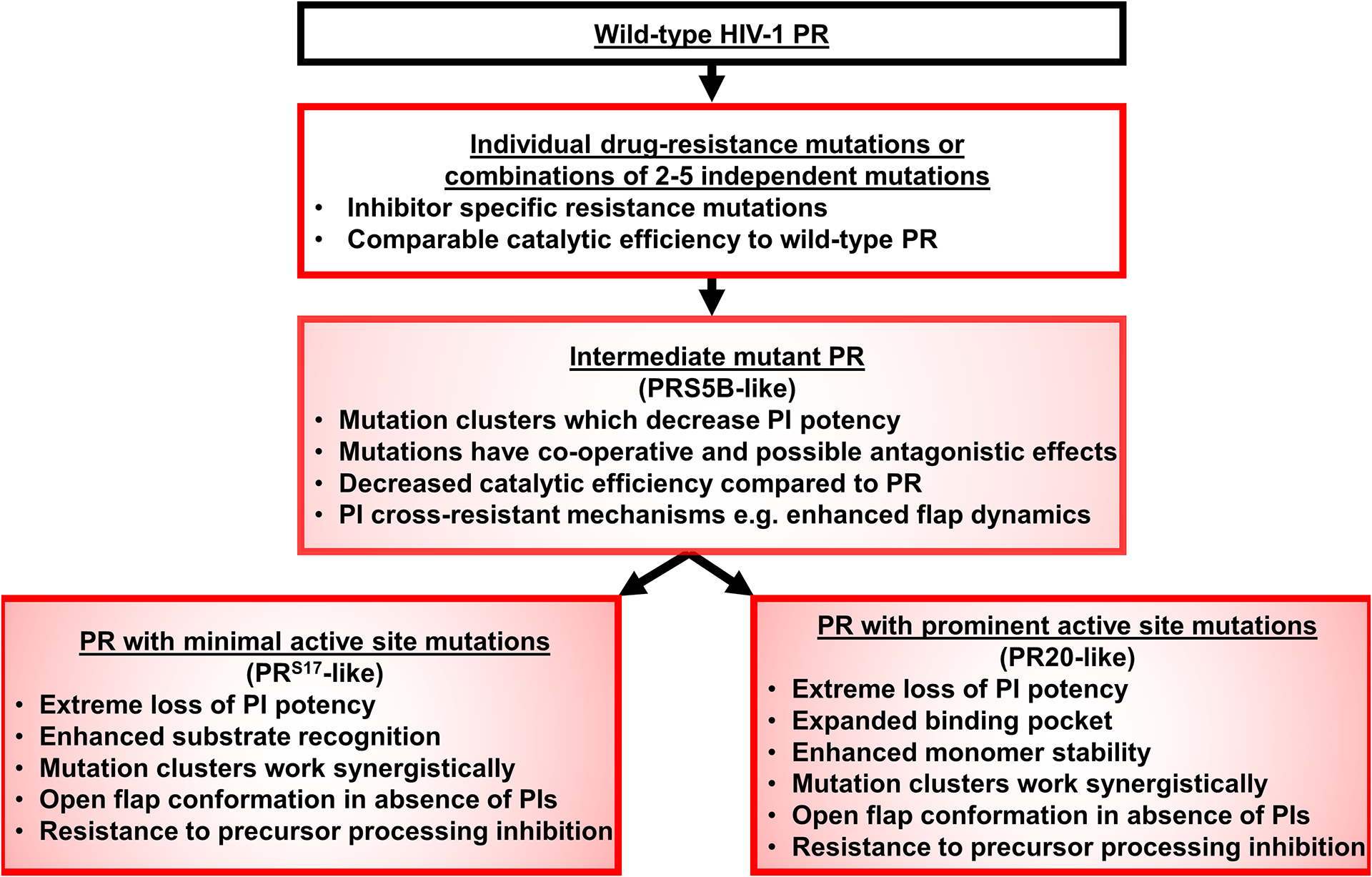
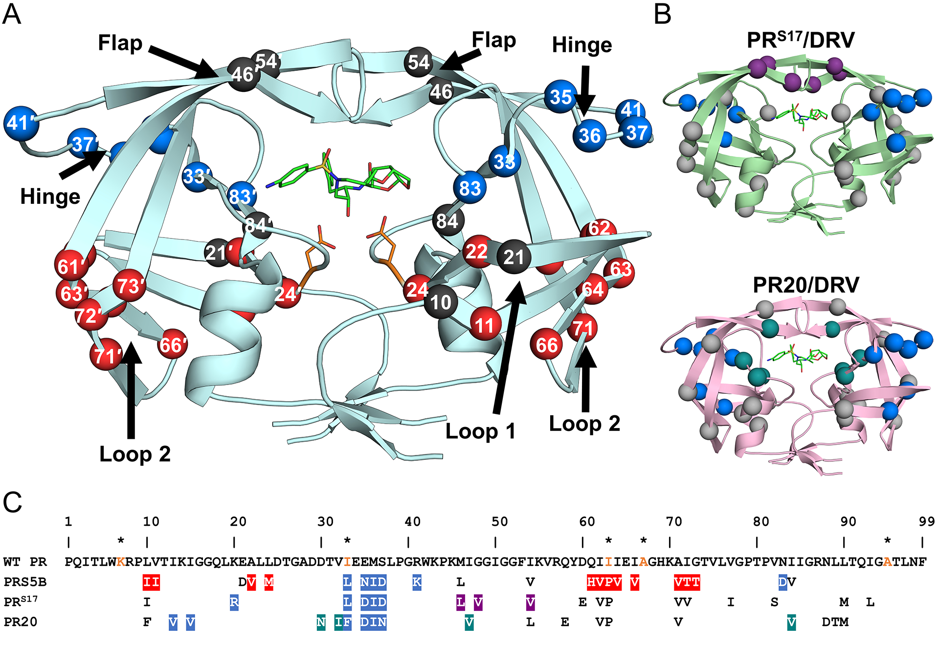
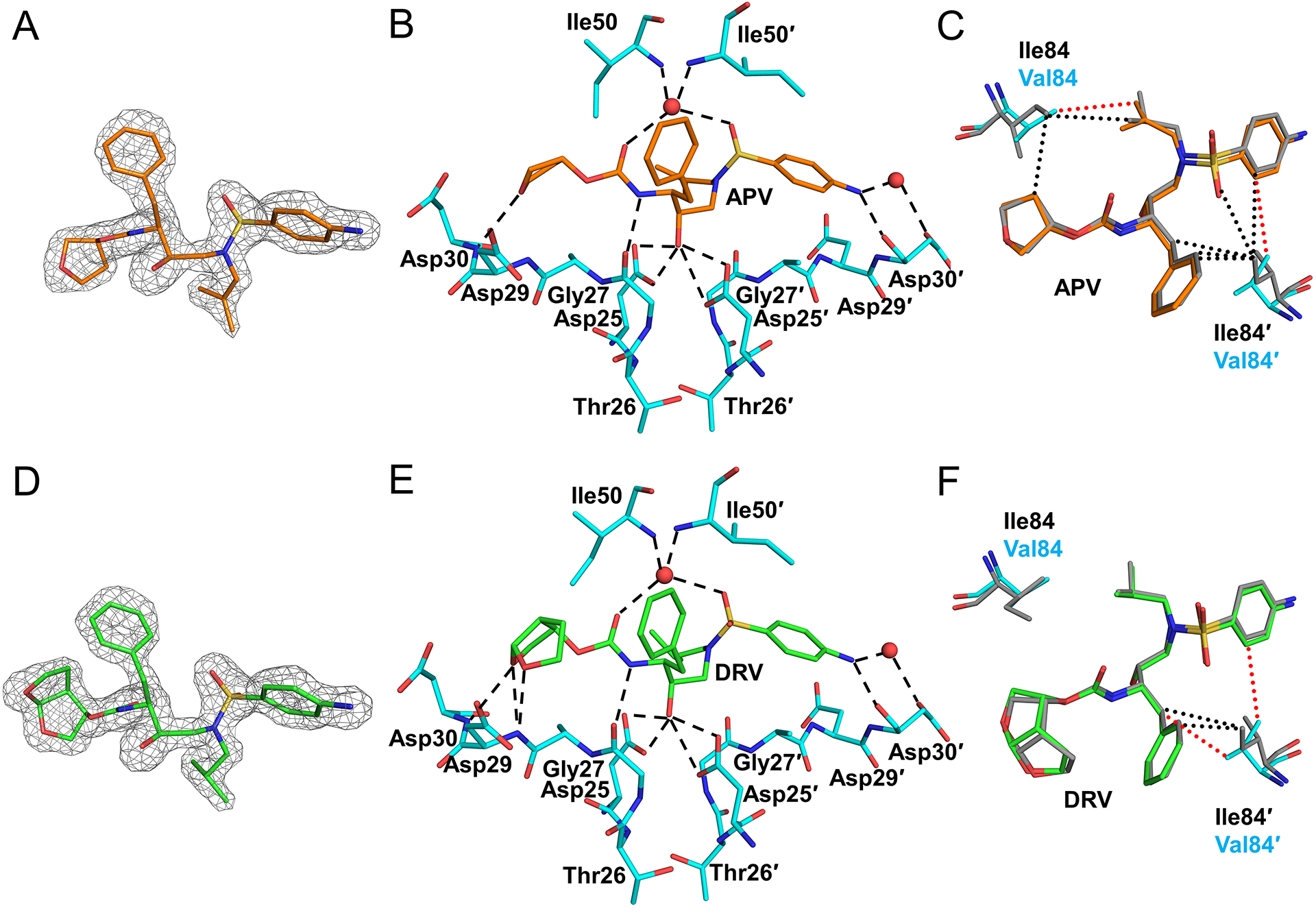
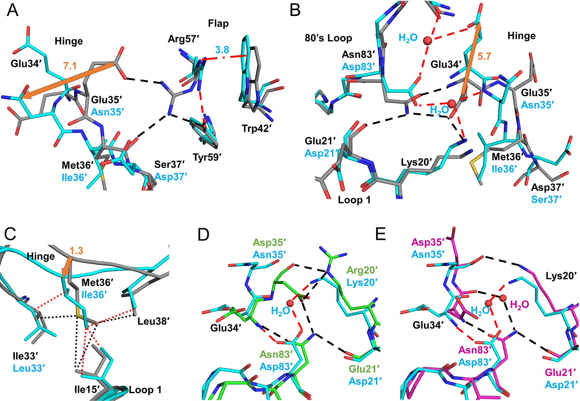
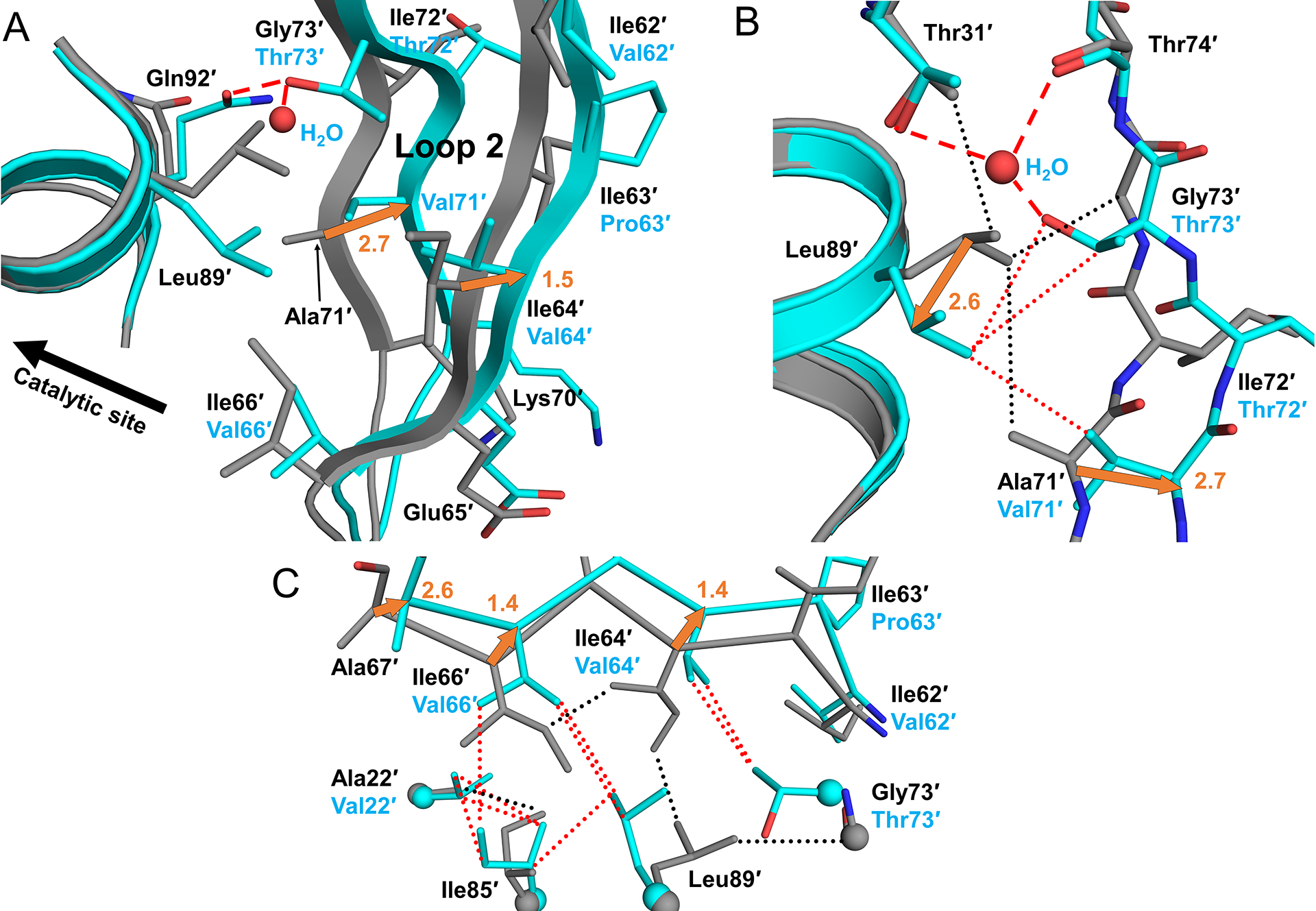
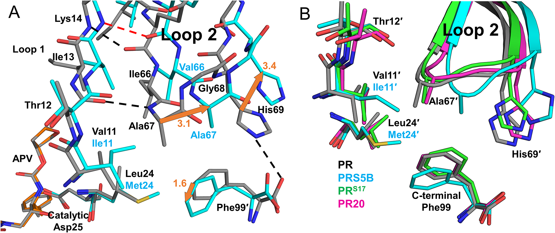
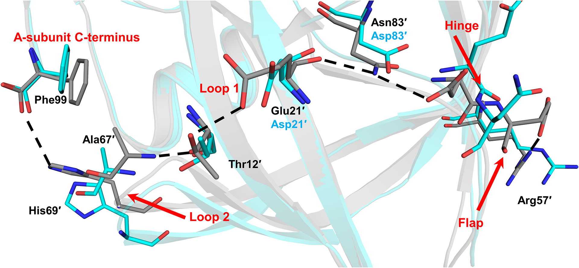
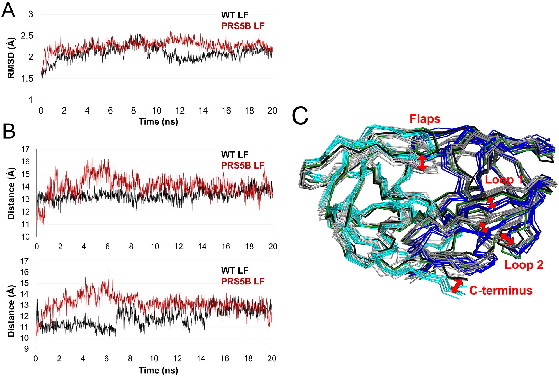
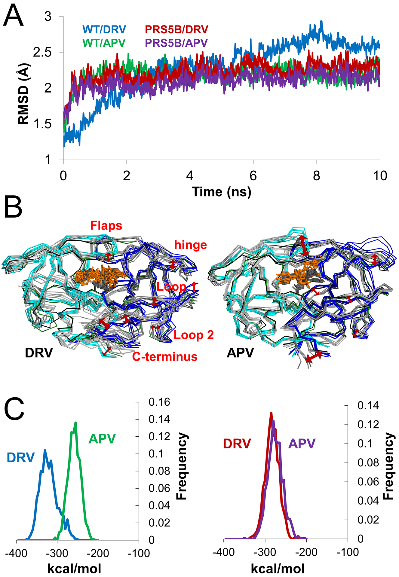
Drug-resistance is a serious problem for treatment of the HIV/AIDS pandemic. Potent clinical inhibitors of HIV-1 protease show several orders of magnitude worse inhibition of highly drug-resistant variants. Hence, the structure and enzyme activities were analyzed for HIV protease mutant HIV-1 protease (EC 3.4.23.16) (PR) with 22 mutations (PRS5B) from a clinical isolate that was selected by machine learning to represent high-level drug-resistance. PRS5B has 22 mutations including only one (I84V) in the inhibitor binding site; however, clinical inhibitors had poor inhibition of PRS5B activity with kinetic inhibition value (K ) values of 4-1000 nm or 18- to 8000-fold worse than for wild-type PR. High-resolution crystal structures of PRS5B complexes with the best inhibitors, amprenavir (APV) and darunavir (DRV) (K ~ 4 nm), revealed only minor changes in protease-inhibitor interactions. Instead, two distinct clusters of mutations in distal regions induce coordinated conformational changes that decrease favorable internal interactions across the entire protein subunit. The largest structural rearrangements are described and compared to other characterized resistant mutants. In the protease hinge region, the N83D mutation eliminates a hydrogen bond connecting the hinge and core of the protease and increases disorder compared to highly resistant mutants PR with 17 mutations and PR with 20 mutations with similar hinge mutations. In a distal β-sheet, mutations G73T and A71V coordinate with accessory mutations to bring about shifts that propagate throughout the subunit. Molecular dynamics simulations of ligand-free dimers show differences consistent with loss of interactions in mutant compared to wild-type PR. Clusters of mutations exhibit both coordinated and antagonistic effects, suggesting PRS5B may represent an intermediate stage in the evolution of more highly resistant variants. DATABASES: Structural data are available in Protein Data Bank under the accession codes 6P9A and 6P9B for PRS5B/DRV and PRS5B/APV, respectively.
耐药性是治疗 HIV/AIDS 大流行的一个严重问题。HIV-1 蛋白酶的强效临床抑制剂对高度耐药变体的抑制作用差几个数量级。因此,对来自临床分离株的 HIV 蛋白酶突变体 HIV-1 蛋白酶(EC 3.4.23.16)(PR)进行了结构和酶活性分析,该分离株通过机器学习选择代表高度耐药性。PRS5B 有 22 个突变,只有一个(I84V)位于抑制剂结合位点;然而,临床抑制剂对 PRS5B 活性的抑制作用很差,动力学抑制值(K )值为 4-1000nm 或比野生型 PR 差 18-8000 倍。PRS5B 与最佳抑制剂安普那韦(APV)和达鲁那韦(DRV)的高分辨率晶体结构复合物显示蛋白酶-抑制剂相互作用只有微小变化。相反,在远端区域的两个不同突变簇诱导协调的构象变化,从而降低整个蛋白质亚基内的有利相互作用。描述并比较了最大的结构重排与其他特征耐药突变体。在蛋白酶铰链区域,N83D 突变消除了连接铰链和蛋白酶核心的氢键,并增加了与具有 17 个突变和具有 20 个突变的高度耐药突变体 PR 相比的无序性,这些突变体具有相似的铰链突变。在远端 β-折叠中,突变 G73T 和 A71V 与辅助突变协同作用,引起贯穿整个亚基的移位。配体自由二聚体的分子动力学模拟显示出与野生型 PR 相比,突变体中相互作用缺失的差异。突变簇表现出协同和拮抗作用,表明 PRS5B 可能代表更高度耐药变体进化的中间阶段。DATABASES:结构数据可在蛋白质数据库中以 6P9A 和 6P9B 的访问代码获得,分别代表 PRS5B/DRV 和 PRS5B/APV。