• 文献检索
  • 文档翻译
  • 深度研究
  • 学术资讯
  • Suppr Zotero 插件Zotero 插件
  • 邀请有礼
  • 套餐&价格
  • 历史记录
应用&插件
Suppr Zotero 插件Zotero 插件浏览器插件Mac 客户端Windows 客户端微信小程序
定价
高级版会员购买积分包购买API积分包
服务
文献检索文档翻译深度研究API 文档MCP 服务
关于我们
关于 Suppr公司介绍联系我们用户协议隐私条款
关注我们

Suppr 超能文献

核心技术专利:CN118964589B侵权必究
粤ICP备2023148730 号-1Suppr @ 2026

文献检索

告别复杂PubMed语法,用中文像聊天一样搜索,搜遍4000万医学文献。AI智能推荐,让科研检索更轻松。

立即免费搜索

文件翻译

保留排版,准确专业,支持PDF/Word/PPT等文件格式,支持 12+语言互译。

免费翻译文档

深度研究

AI帮你快速写综述,25分钟生成高质量综述,智能提取关键信息,辅助科研写作。

立即免费体验

微计算机断层扫描与锥形束计算机断层扫描在骨质量评估中的比较以及白蛋白浸渍同种异体骨移植上颌窦增强后的长期容积研究。

Comparison between Micro-Computed Tomography and Cone-Beam Computed Tomography in the Assessment of Bone Quality and a Long-Term Volumetric Study of the Augmented Sinus Grafted with an Albumin Impregnated Allograft.

作者信息

Kivovics Márton, Szabó Bence Tamás, Németh Orsolya, Iványi Dóra, Trimmel Bálint, Szmirnova Ilona, Orhan Kaan, Mijiritsky Eitan, Szabó György, Dobó-Nagy Csaba

机构信息

Department of Community Dentistry, Semmelweis University, 1088 Budapest, Hungary.

Department of Oral Diagnostics, Semmelweis University, 1088 Budapest, Hungary.

出版信息

J Clin Med. 2020 Jan 21;9(2):303. doi: 10.3390/jcm9020303.

DOI:10.3390/jcm9020303
PMID:31973237
原文链接:https://pmc.ncbi.nlm.nih.gov/articles/PMC7073646/
Abstract

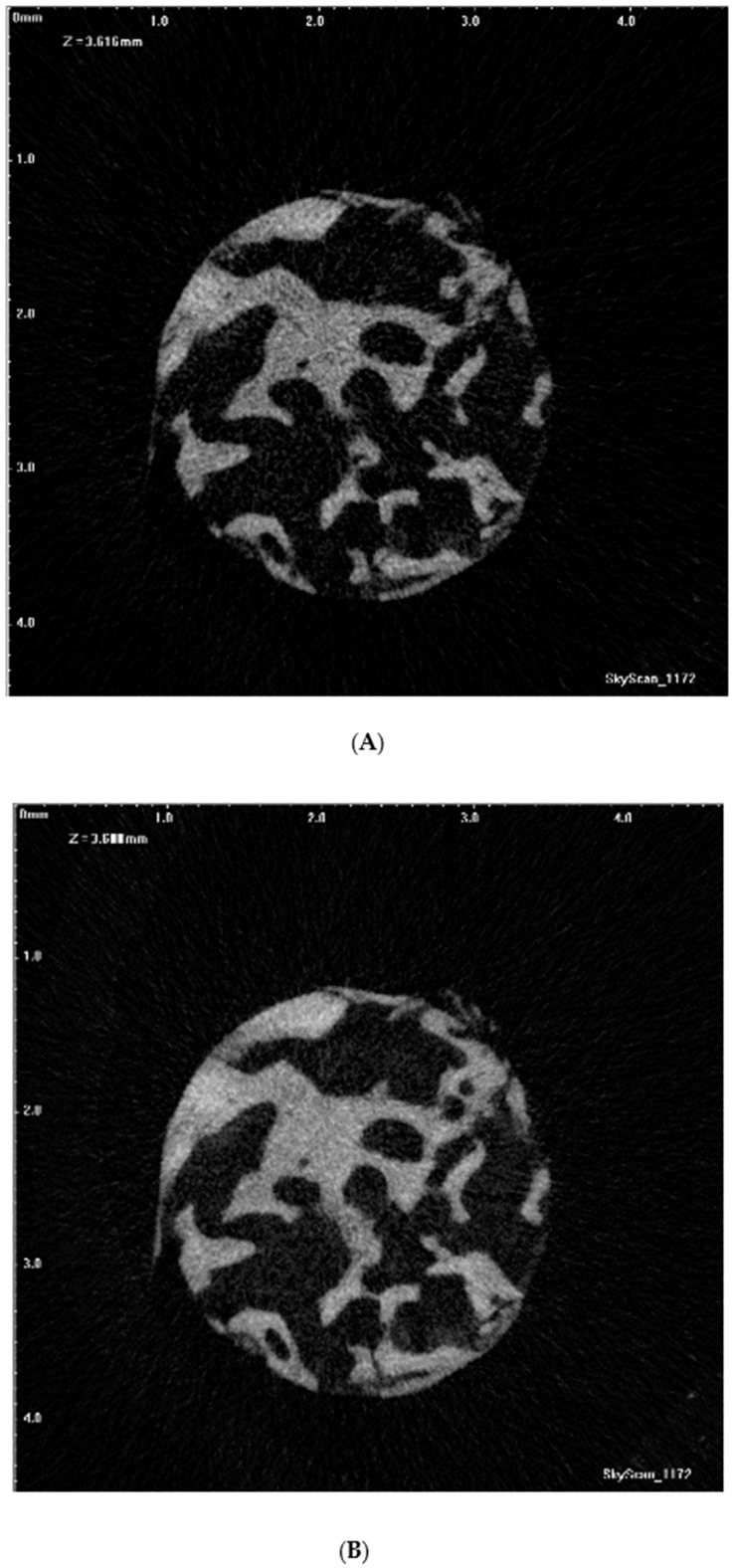
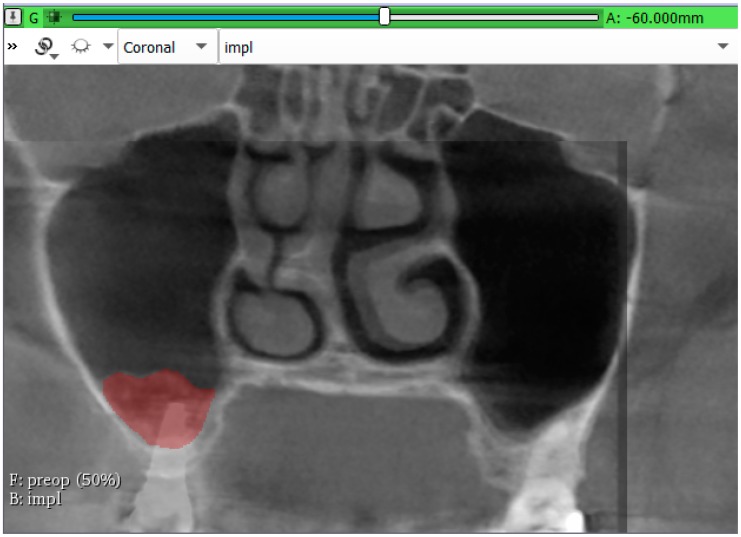
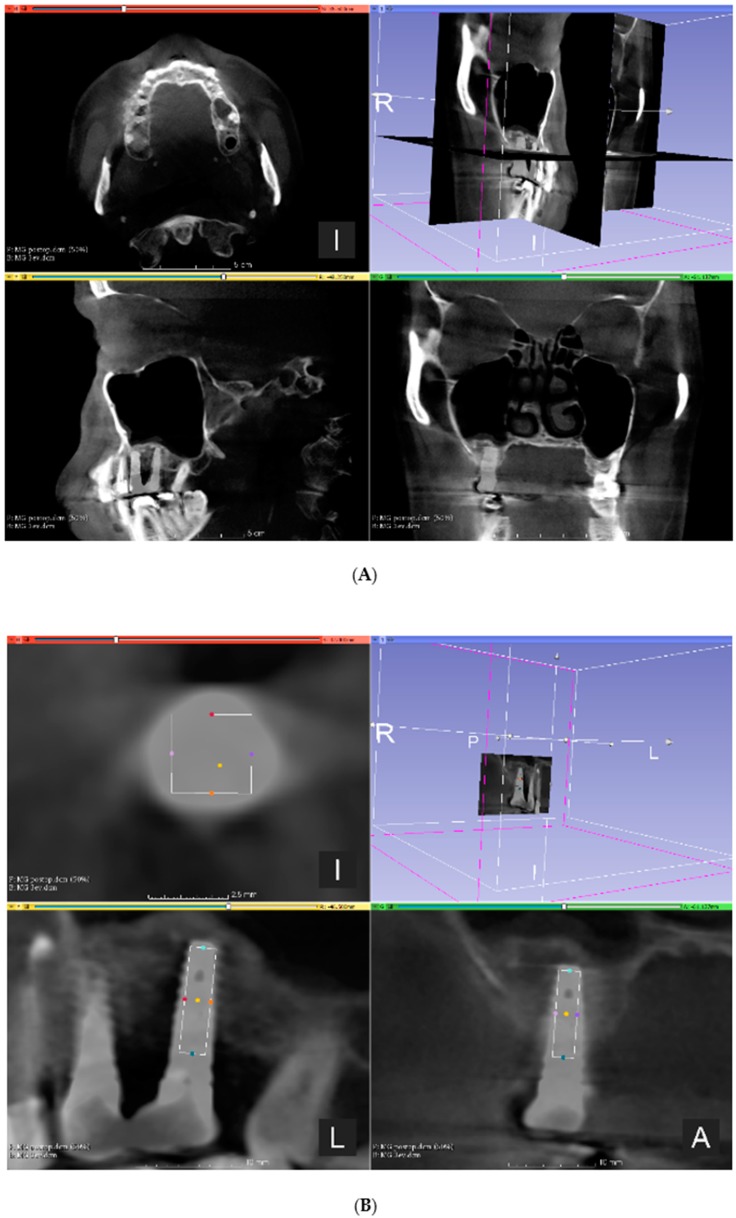
The purpose of our study was to compare micromorphometric data obtained by cone-beam computed-tomography (CBCT) and microcomputed-tomography (micro-CT) of the augmented sinus and to evaluate the long-term stability of the bone gain achieved using BoneAlbumin. Sinus lifts, and after 6-months, healing bone-biopsy and implant placement were carried out. Specimens were analyzed by micro-CT. A total of 16 samples were collected from nine patients (mean age 54.7 ± 6.5 years). Pre-, postoperative, and 3-year control CBCT-data were registered to determine from where the biopsy samples were harvested. Micromorphometric variables were calculated from the micro-CT- and CBCT-data, and their correlation was determined by Spearman's test. The volume of augmented bone was calculated at the time of implant placement and after 3 years. A positive correlation was found between bone-volume fraction, trabecular-separation, open-, and total-porosity, while a negative correlation was found between trabecular-thickness obtained from CBCT- and micro-CT-data ( < 0.05). Mean volumetric reduction of 39.28% (11.88-60.02%) was observed. Correlation of CBCT- and micro-CT-data suggested that micromorphometric analysis of CBCT reconstructions of the augmented sinuses provided reliable information on the microarchitecture of augmented bone. CBCT as a modality might be adequate in the analysis of bone quality in the augmented sinus. At the 3-year, control sinus grafts showed volumetric stability.

摘要

我们研究的目的是比较通过锥形束计算机断层扫描(CBCT)和显微计算机断层扫描(micro-CT)获得的上颌窦增高术的微观形态测量数据,并评估使用骨白蛋白实现的骨增量的长期稳定性。进行上颌窦提升术,6个月后进行愈合骨活检和种植体植入。通过micro-CT分析样本。共从9名患者(平均年龄54.7±6.5岁)中收集了16个样本。记录术前、术后和3年对照CBCT数据,以确定活检样本的采集位置。根据micro-CT和CBCT数据计算微观形态测量变量,并通过Spearman检验确定它们之间的相关性。在种植体植入时和3年后计算增高骨的体积。发现骨体积分数、小梁间距、开孔率和总孔隙率之间呈正相关,而从CBCT和micro-CT数据获得的小梁厚度之间呈负相关(<0.05)。观察到平均体积减少39.28%(11.88 - 60.02%)。CBCT和micro-CT数据的相关性表明,对上颌窦增高术的CBCT重建进行微观形态测量分析可为增高骨的微观结构提供可靠信息。CBCT作为一种方法在分析上颌窦增高术中的骨质量方面可能是足够的。在3年时,对照鼻窦移植物显示出体积稳定性。

https://cdn.ncbi.nlm.nih.gov/pmc/blobs/b081/7073646/26bef626b508/jcm-09-00303-g003.jpg
https://cdn.ncbi.nlm.nih.gov/pmc/blobs/b081/7073646/666c83ba95e5/jcm-09-00303-g001.jpg
https://cdn.ncbi.nlm.nih.gov/pmc/blobs/b081/7073646/a44615364743/jcm-09-00303-g002.jpg
https://cdn.ncbi.nlm.nih.gov/pmc/blobs/b081/7073646/26bef626b508/jcm-09-00303-g003.jpg
https://cdn.ncbi.nlm.nih.gov/pmc/blobs/b081/7073646/666c83ba95e5/jcm-09-00303-g001.jpg
https://cdn.ncbi.nlm.nih.gov/pmc/blobs/b081/7073646/a44615364743/jcm-09-00303-g002.jpg
https://cdn.ncbi.nlm.nih.gov/pmc/blobs/b081/7073646/26bef626b508/jcm-09-00303-g003.jpg

相似文献

1
Comparison between Micro-Computed Tomography and Cone-Beam Computed Tomography in the Assessment of Bone Quality and a Long-Term Volumetric Study of the Augmented Sinus Grafted with an Albumin Impregnated Allograft.微计算机断层扫描与锥形束计算机断层扫描在骨质量评估中的比较以及白蛋白浸渍同种异体骨移植上颌窦增强后的长期容积研究。
J Clin Med. 2020 Jan 21;9(2):303. doi: 10.3390/jcm9020303.
2
The reliability of cone-beam computed tomography to assess bone density at dental implant recipient sites: a histomorphometric analysis by micro-CT.锥形束计算机断层扫描评估种植体接受部位骨密度的可靠性:微 CT 组织形态计量学分析。
Clin Oral Implants Res. 2013 Aug;24(8):871-9. doi: 10.1111/j.1600-0501.2011.02390.x. Epub 2012 Jan 17.
3
Bone quality evaluation at dental implant site using multislice CT, micro-CT, and cone beam CT.使用多层螺旋CT、显微CT和锥形束CT对牙种植部位进行骨质量评估。
Clin Oral Implants Res. 2015;26(1):e1-7. doi: 10.1111/clr.12315. Epub 2013 Dec 11.
4
The Accuracy of Cone-Beam Computed Tomography for Evaluating Bone Density and Cortical Bone Thickness at the Implant Site: Micro-Computed Tomography and Histologic Analysis.锥形束计算机断层扫描评估种植部位骨密度和皮质骨厚度的准确性:显微计算机断层扫描和组织学分析
J Craniofac Surg. 2018 Nov;29(8):2026-2031. doi: 10.1097/SCS.0000000000004672.
5
Implant-guided volumetric analysis of edentulous maxillary bone with cone-beam computerized tomography scan. Maxillary sinus pneumatization classification.锥形束计算机断层扫描对上颌无牙颌骨进行种植引导的容积分析。上颌窦气化分类。
J Oral Implantol. 2012 Aug;38(4):377-90. doi: 10.1563/AAID-JOI-D-11-00212.
6
Evaluation of maxillary trabecular microstructure as an indicator of implant stability by using 2 cone beam computed tomography systems and micro-computed tomography.使用 2 种锥形束 CT 系统和微计算机断层扫描评估上颌窦小梁微结构作为种植体稳定性的指标。
Oral Surg Oral Med Oral Pathol Oral Radiol. 2019 Mar;127(3):247-256. doi: 10.1016/j.oooo.2018.11.014. Epub 2018 Dec 1.
7
The assessment of trabecular bone parameters and cortical bone strength: a comparison of micro-CT and dental cone-beam CT.小梁骨参数和皮质骨强度评估:微 CT 和口腔锥形束 CT 的比较。
J Biomech. 2013 Oct 18;46(15):2611-8. doi: 10.1016/j.jbiomech.2013.08.004. Epub 2013 Aug 24.
8
Effect of Scanning Resolution on the Prediction of Trabecular Bone Microarchitectures Using Dental Cone Beam Computed Tomography.扫描分辨率对使用牙科锥形束计算机断层扫描预测小梁骨微结构的影响。
Diagnostics (Basel). 2020 Jun 3;10(6):368. doi: 10.3390/diagnostics10060368.
9
Improving the prediction of the trabecular bone microarchitectural parameters using dental cone-beam computed tomography.使用牙科锥形束计算机断层扫描改善小梁骨微结构参数的预测。
BMC Med Imaging. 2019 Jan 23;19(1):10. doi: 10.1186/s12880-019-0313-9.
10
[Maxillary sinus floor augmentation with Bio-Oss as a sole graft: a histologic, radiographic and preliminary clinical study].[使用Bio-Oss作为单一移植物进行上颌窦底提升:组织学、影像学及初步临床研究]
Zhonghua Kou Qiang Yi Xue Za Zhi. 2013 Sep;48(9):513-7.

引用本文的文献

1
Fractal Analysis as a Predictor of Early Implant Loss: A Retrospective Study.分形分析作为早期种植体丢失的预测指标:一项回顾性研究。
Int Dent J. 2025 Aug 30;75(6):103880. doi: 10.1016/j.identj.2025.103880.
2
A systematic review and meta-analysis on the concept of bone quality in dento-maxillofacial Cone Beam Computed Tomography.关于牙颌面锥形束计算机断层扫描中骨质量概念的系统评价与荟萃分析。
Radiol Med. 2025 Jul 16. doi: 10.1007/s11547-025-02052-5.
3
Cone beam computed tomography vs. microcomputed tomography for trabecular bone healing assessment : An ex vivo study in rabbit mandibles.

本文引用的文献

1
The micro-CT evaluation of enamel-cement thickness, abrasion, and mineral density in teeth in the postmortem interval (PMI): new parameters for the determination of PMI.死后间隔时间内(PMI)牙齿釉牙骨质界厚度、磨损和矿物质密度的微 CT 评估:PMI 测定的新参数。
Int J Legal Med. 2020 Mar;134(2):645-653. doi: 10.1007/s00414-019-02104-2. Epub 2019 Jul 3.
2
Medical Treatment for Osteoporosis: From Molecular to Clinical Opinions.骨质疏松症的治疗:从分子到临床观点。
Int J Mol Sci. 2019 May 6;20(9):2213. doi: 10.3390/ijms20092213.
3
Effectiveness of Platelet-Rich Fibrin as an Adjunctive Material to Bone Graft in Maxillary Sinus Augmentation: A Meta-Analysis of Randomized Controlled Trails.
锥形束计算机断层扫描与微型计算机断层扫描用于小梁骨愈合评估的比较:兔下颌骨的体外研究
J Orofac Orthop. 2025 May 20. doi: 10.1007/s00056-025-00589-8.
4
Bone Densitometric Analysis using Cone Beam Computed Tomography (Cone Beam CT) and Computed Tomography (CT): Establishing the Correlation between Predicted and Actual Values.使用锥形束计算机断层扫描(锥形束CT)和计算机断层扫描(CT)进行骨密度分析:建立预测值与实际值之间的相关性。
Adv Biomed Res. 2025 Jan 30;14:8. doi: 10.4103/abr.abr_313_24. eCollection 2025.
5
Prevalence of middle mesial root canal in mandibular molars in an Iranian population: A micro-computed tomography study.伊朗人群下颌磨牙近中中根管的患病率:一项显微计算机断层扫描研究。
Dent Res J (Isfahan). 2024 Jan 25;21:9. eCollection 2024.
6
Are Artificial Intelligence-Assisted Three-Dimensional Histological Reconstructions Reliable for the Assessment of Trabecular Microarchitecture?人工智能辅助的三维组织学重建对小梁微结构评估是否可靠?
J Clin Med. 2024 Feb 15;13(4):1106. doi: 10.3390/jcm13041106.
7
Correlation between gray values in cone-beam computed tomography and histomorphometric analysis.锥形束计算机断层扫描中的灰度值与组织形态计量学分析之间的相关性。
Imaging Sci Dent. 2022 Dec;52(4):375-382. doi: 10.5624/isd.20220051. Epub 2022 Oct 12.
8
The Use of CBCT in Evaluating the Health and Pathology of the Maxillary Sinus.锥形束计算机断层扫描(CBCT)在上颌窦健康与病变评估中的应用
Diagnostics (Basel). 2022 Nov 16;12(11):2819. doi: 10.3390/diagnostics12112819.
9
Criteria for the Classification of the Interradicular Septum Shape in Maxillary Molars with Clinical Importance for Prosthetic-Driven Immediate Implant Placement.上颌磨牙根间间隔形态分类标准及其对修复驱动即刻种植植入的临床意义
Diagnostics (Basel). 2022 Jun 10;12(6):1432. doi: 10.3390/diagnostics12061432.
10
Micro-computed tomography in preventive and restorative dental research: A review.微计算机断层扫描在口腔预防与修复研究中的应用综述
Imaging Sci Dent. 2021 Dec;51(4):341-350. doi: 10.5624/isd.20210087. Epub 2021 Oct 15.
富血小板纤维蛋白作为上颌窦提升中骨移植辅助材料的有效性:随机对照试验的荟萃分析。
Biomed Res Int. 2019 Mar 17;2019:7267062. doi: 10.1155/2019/7267062. eCollection 2019.
4
Microarchitecture of the Augmented Bone Following Sinus Elevation with an Albumin Impregnated Demineralized Freeze-Dried Bone Allograft (BoneAlbumin) versus Anorganic Bovine Bone Mineral: A Randomized Prospective Clinical, Histomorphometric, and Micro-Computed Tomography Study.白蛋白浸渍脱矿冻干骨移植材料(BoneAlbumin)与无机牛骨矿物质用于上颌窦提升术后增强骨的微观结构:一项随机前瞻性临床、组织形态计量学及微型计算机断层扫描研究
Materials (Basel). 2018 Jan 28;11(2):202. doi: 10.3390/ma11020202.
5
Mandibular Bone Structure Analysis Using Cone Beam Computed Tomography vs Primary Implant Stability: An Ex Vivo Study.使用锥形束计算机断层扫描分析下颌骨结构与种植体初期稳定性的体外研究
Int J Oral Maxillofac Implants. 2017 Nov/Dec;32(6):1257-1265. doi: 10.11607/jomi.6210.
6
Current bone substitutes for implant dentistry.目前用于种植牙科的骨替代物。
J Prosthodont Res. 2018 Apr;62(2):152-161. doi: 10.1016/j.jpor.2017.08.010. Epub 2017 Sep 15.
7
Accuracy of Implant Placement with Computer-Guided Surgery: A Systematic Review and Meta-Analysis Comparing Cadaver, Clinical, and In Vitro Studies.计算机辅助手术中种植体植入的准确性:一项比较尸体研究、临床研究和体外研究的系统评价与荟萃分析
Int J Oral Maxillofac Implants. 2018 January/February;33(1):101–115. doi: 10.11607/jomi.5556. Epub 2017 Jun 20.
8
Accuracy and reliability of different cone beam computed tomography (CBCT) devices for structural analysis of alveolar bone in comparison with multislice CT and micro-CT.与多层螺旋CT和微型CT相比,不同锥形束计算机断层扫描(CBCT)设备用于牙槽骨结构分析的准确性和可靠性。
Eur J Oral Implantol. 2017;10(1):95-105.
9
Efficacy of Cone-Beam Computed Tomography in Evaluating Bone Quality for Optimum Implant Treatment Planning.锥形束计算机断层扫描在评估骨质量以优化种植体治疗规划中的疗效。
Implant Dent. 2017 Jun;26(3):405-411. doi: 10.1097/ID.0000000000000542.
10
Microarchitectural study of the augmented bone following ridge preservation with a porcine xenograft and a collagen membrane: preliminary report of a prospective clinical, histological, and micro-computed tomography analysis.使用猪异种移植物和胶原膜进行牙槽嵴保存后植骨增强的微观结构研究:一项前瞻性临床、组织学和显微计算机断层扫描分析的初步报告
Int J Oral Maxillofac Surg. 2017 Feb;46(2):250-260. doi: 10.1016/j.ijom.2016.10.010. Epub 2016 Nov 8.