Center for the Developing Brain, Children's National Hospital, Washington, DC.
Department of Biostatistics and Study Methodology, Children's Research Institute, Children's National Hospital, Washington, DC.
JAMA Netw Open. 2020 Jan 3;3(1):e1919940. doi: 10.1001/jamanetworkopen.2019.19940.
Prenatal maternal stress is increasingly associated with adverse outcomes in pregnant women and their offspring. However, the association between maternal stress and human fetal brain growth and metabolism is unknown.
To identify the association between prenatal maternal psychological distress and fetal brain growth, cortical maturation, and biochemical development using advanced 3-dimensional volumetric magnetic resonance imaging (MRI) and proton magnetic resonance spectroscopy (1H-MRS).
DESIGN, SETTING, AND PARTICIPANTS: This cohort study prospectively recruited pregnant women from low-risk obstetric clinics in Washington, DC, from January 1, 2016, to April 17, 2019. Participants were healthy volunteers with a normal prenatal medical history, no chronic or pregnancy-induced physical or mental illnesses, and normal results on fetal ultrasonography and biometry studies. Fetal brain MRI studies were performed at 2 time points between 24 and 40 weeks' gestation.
Prenatal maternal stress, anxiety, and depression.
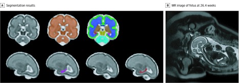
Volumes of fetal total brain, cortical gray matter, white matter, deep gray matter, cerebellum, brainstem, and hippocampus were measured from 3-dimensional reconstructed T2-weighted MRI scans. Cortical folding measurements included local gyrification index, sulcal depth, and curvedness. Fetal brain N-acetylaspartate, creatine, and choline levels were quantified using 1H-MRS. Maternal stress, depression, and anxiety were measured with the Perceived Stress Scale (PSS), Edinburgh Postnatal Depression Scale (EPDS), Spielberger State Anxiety Inventory (SSAI), and Spielberger Trait Anxiety Inventory (STAI).
A total of 193 MRI studies were performed in 119 pregnant women (67 [56%] carrying male fetuses and 52 [44%], female fetuses; maternal mean [SD] age, 34.46 [5.95] years) between 24 and 40 gestational weeks. All women were high school graduates, 99 (83%) were college graduates, and 100 (84%) reported professional employment. Thirty-two women (27%) had positive scores for stress, 31 (26%) for anxiety, and 13 (11%) for depression. Maternal trait anxiety was associated with smaller fetal left hippocampal volume (STAI score: -0.002 cm3; 95% CI, -0.003 to -0.0008 cm3; P = .004). Maternal anxiety and stress were associated with increased fetal cortical gyrification in the frontal lobe (β for SSAI score: 0.004 [95% CI, 0.001-0.006; P = .002]; β for STAI score: 0.004 [95% CI, 0.002-0.006; P < .001]; β for PSS score: 0.005 [95% CI, 0.001-0.008; P = .005]) and temporal lobe (β for SSAI score: 0.004 [95% CI, 0.001-0.007; P = .004]; β for STAI score: 0.004 [95% CI, 0.0008-0.006; P = .01]). Elevated maternal depression was associated with decreased creatine (EPDS score: -0.04; 95% CI, -0.06 to -0.02; P = .005) and choline (EPDS score: -0.03; 95% CI, -0.05 to -0.01; P = .02) levels in the fetal brain.
This study found that the prevalence of maternal psychological distress in healthy, well-educated, and employed pregnant women was high, underappreciated, and associated with impaired fetal brain biochemistry and hippocampal growth as well as accelerated cortical folding. These findings appear to support the need for routine mental health surveillance for all pregnant women and targeted interventions in women with elevated psychological distress.
产前母体压力与孕妇及其后代的不良结局日益相关。然而,母体压力与人类胎儿大脑生长和代谢之间的关系尚不清楚。
使用先进的三维容积磁共振成像(MRI)和质子磁共振波谱(1H-MRS)技术,确定产前母体心理困扰与胎儿大脑生长、皮质成熟和生化发育之间的关系。
设计、地点和参与者:本队列研究前瞻性地招募了来自华盛顿特区低风险产科诊所的健康孕妇志愿者,招募时间为 2016 年 1 月 1 日至 2019 年 4 月 17 日。参与者为具有正常产前病史、无慢性或妊娠引起的身体或精神疾病以及正常胎儿超声和生物测量研究结果的健康志愿者。在妊娠 24 至 40 周之间的两个时间点进行胎儿脑 MRI 研究。
产前母体压力、焦虑和抑郁。
从三维重建 T2 加权 MRI 扫描中测量胎儿全脑、皮质灰质、白质、深部灰质、小脑、脑干和海马体的体积。皮质折叠测量包括局部脑回指数、脑沟深度和弯曲度。使用 1H-MRS 量化胎儿脑内 N-乙酰天冬氨酸、肌酸和胆碱水平。使用感知压力量表(PSS)、爱丁堡产后抑郁量表(EPDS)、斯皮尔伯格状态焦虑量表(SSAI)和斯皮尔伯格特质焦虑量表(STAI)测量母体压力、抑郁和焦虑。
在妊娠 24 至 40 周之间,对 119 名孕妇(67 名携带男性胎儿,52 名携带女性胎儿;母亲平均[标准差]年龄 34.46[5.95]岁)的 193 项 MRI 研究进行了分析。所有女性均为高中毕业生,99 人(83%)为大学毕业生,100 人(84%)有专业就业。32 名女性(27%)的压力评分阳性,31 名(26%)的焦虑评分阳性,13 名(11%)的抑郁评分阳性。母体特质焦虑与左侧海马体体积减小相关(STAI 评分:-0.002cm3;95%CI,-0.003 至 -0.0008cm3;P=0.004)。母体焦虑和压力与额叶皮质脑回增加有关(SSAI 评分的β值:0.004[95%CI,0.001 至 0.006;P=0.002];STAI 评分的β值:0.004[95%CI,0.002 至 0.006;P<0.001];PSS 评分的β值:0.005[95%CI,0.001 至 0.008;P=0.005])和颞叶(SSAI 评分的β值:0.004[95%CI,0.001 至 0.007;P=0.004];STAI 评分的β值:0.004[95%CI,0.0008 至 0.006;P=0.01])。母体抑郁评分升高与胎儿脑内肌酸(EPDS 评分:-0.04;95%CI,-0.06 至 -0.02;P=0.005)和胆碱(EPDS 评分:-0.03;95%CI,-0.05 至 -0.01;P=0.02)水平降低有关。
本研究发现,在健康、受过良好教育和有职业的孕妇中,母体心理困扰的患病率较高,且未得到充分认识,与胎儿脑生物化学和海马体生长受损以及皮质折叠加速有关。这些发现似乎支持对所有孕妇进行常规心理健康监测和对有较高心理困扰的女性进行有针对性的干预。