Developing Brain Institute, Children's National Hospital, Washington, DC.
Division of Neonatology, Children's National Hospital, Washington, DC.
JAMA Netw Open. 2022 Apr 1;5(4):e229244. doi: 10.1001/jamanetworkopen.2022.9244.
Prenatal maternal psychological distress is associated with disturbances in fetal brain development. However, the association between altered fetal brain development, prenatal maternal psychological distress, and long-term neurodevelopmental outcomes is unknown.
To determine the association of fetal brain development using 3-dimensional magnetic resonance imaging (MRI) volumes, cortical folding, and metabolites in the setting of maternal psychological distress with infant 18-month neurodevelopment.
DESIGN, SETTING, AND PARTICIPANTS: Healthy mother-infant dyads were prospectively recruited into a longitudinal observational cohort study from January 2016 to October 2020 at Children's National Hospital in Washington, DC. Data analysis was performed from January 2016 to July 2021.
Prenatal maternal stress, anxiety, and depression.
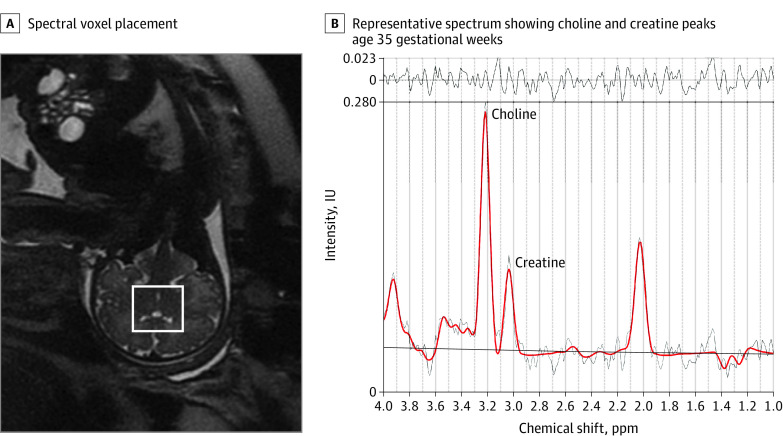
Prenatal maternal stress, anxiety, and depression were measured using validated self-report questionnaires. Fetal brain volumes and cortical folding were measured from 3-dimensional, reconstructed T2-weighted MRI scans. Fetal brain creatine and choline were quantified using proton magnetic resonance spectroscopy. Infant neurodevelopment at 18 months was measured using Bayley Scales of Infant and Toddler Development III and Infant-Toddler Social and Emotional Assessment. The parenting stress in the parent-child dyad was measured using the Parenting Stress Index-Short Form at 18-month testing.
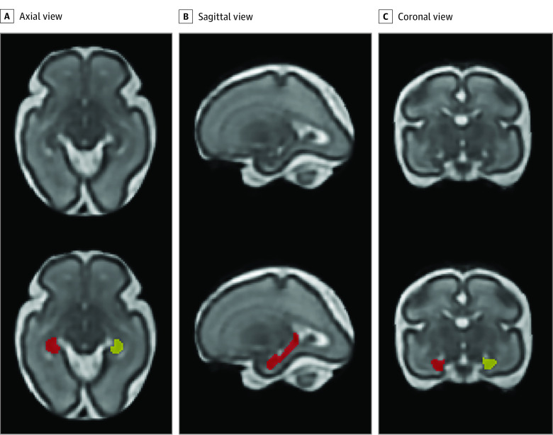
The cohort consisted of 97 mother-infant dyads (mean [SD] maternal age, 34.79 [5.64] years) who underwent 184 fetal MRI visits (87 participants with 2 fetal studies each) with maternal psychological distress measures between 24 and 40 gestational weeks and completed follow-up infant neurodevelopmental testing. Prenatal maternal stress was negatively associated with infant cognitive performance (β = -0.51; 95% CI, -0.92 to -0.09; P = .01), and this association was mediated by fetal left hippocampal volume. In addition, prenatal maternal anxiety, stress, and depression were positively associated with all parenting stress measures at 18-month testing. Finally, fetal cortical local gyrification index and sulcal depth were negatively associated with infant social-emotional performance (local gyrification index: β = -54.62; 95% CI, -85.05 to -24.19; P < .001; sulcal depth: β = -14.22; 95% CI, -23.59 to -4.85; P = .002) and competence scores (local gyrification index: β = -24.01; 95% CI, -40.34 to -7.69; P = .003; sulcal depth: β = -7.53; 95% CI, -11.73 to -3.32; P < .001).
In this cohort study of 97 mother-infant dyads, fetal cortical local gyrification index and sulcal depth were associated with infant 18-month social-emotional and competence outcomes, and fetal left hippocampal volume mediated the association between prenatal maternal stress and infant cognitive outcome. These findings suggest that altered prenatal brain development in the setting of elevated maternal distress has adverse infant sociocognitive outcomes, and identifying early biomarkers associated with long-term neurodevelopment may assist in early targeted interventions.
产前母体心理困扰与胎儿大脑发育障碍有关。然而,胎儿大脑发育的改变、产前母体心理困扰与长期神经发育结果之间的关联尚不清楚。
使用 3 维磁共振成像(MRI)体积、皮质折叠和代谢物来确定胎儿大脑发育与母体心理困扰情况下婴儿 18 个月神经发育的关系。
设计、设置和参与者:健康母婴对从 2016 年 1 月至 2020 年 10 月在华盛顿特区儿童国家医院前瞻性招募入纵向观察队列研究。数据分析于 2016 年 1 月至 2021 年 7 月进行。
产前母亲的压力、焦虑和抑郁。
使用经过验证的自我报告问卷测量产前母亲的压力、焦虑和抑郁。从 3 维重建 T2 加权 MRI 扫描测量胎儿大脑体积和皮质折叠。使用质子磁共振波谱定量胎儿脑内肌酸和胆碱。婴儿 18 个月的神经发育采用贝利婴幼儿发展量表第三版和婴儿-幼儿社会和情感评估进行测量。在 18 个月的测试中,使用父母-子女关系中的父母压力指数-简短形式来测量父母在父母-子女关系中的压力。
该队列包括 97 对母婴(平均[SD]母亲年龄,34.79[5.64]岁),他们接受了 184 次胎儿 MRI 检查(87 名参与者各有 2 次胎儿研究),在妊娠 24 至 40 周时进行了母亲心理困扰测量,并完成了后续的婴儿神经发育测试。产前母亲压力与婴儿认知表现呈负相关(β=-0.51;95%CI,-0.92 至-0.09;P=0.01),这种关联通过胎儿左海马体体积来介导。此外,产前母亲的焦虑、压力和抑郁与 18 个月测试时的所有育儿压力测量均呈正相关。最后,胎儿皮质局部脑回指数和脑沟深度与婴儿社会情感表现呈负相关(局部脑回指数:β=-54.62;95%CI,-85.05 至-24.19;P<0.001;脑沟深度:β=-14.22;95%CI,-23.59 至-4.85;P=0.002)和能力评分(局部脑回指数:β=-24.01;95%CI,-40.34 至-7.69;P=0.003;脑沟深度:β=-7.53;95%CI,-11.73 至-3.32;P<0.001)。
在这项针对 97 对母婴的队列研究中,胎儿皮质局部脑回指数和脑沟深度与婴儿 18 个月的社会情感和能力结果相关,而胎儿左海马体体积则介导了产前母亲压力与婴儿认知结果之间的关联。这些发现表明,在母体压力升高的情况下,胎儿大脑发育的改变与婴儿的社会认知结果不良有关,识别与长期神经发育相关的早期生物标志物可能有助于早期的针对性干预。