• 文献检索
  • 文档翻译
  • 深度研究
  • 学术资讯
  • Suppr Zotero 插件Zotero 插件
  • 邀请有礼
  • 套餐&价格
  • 历史记录
应用&插件
Suppr Zotero 插件Zotero 插件浏览器插件Mac 客户端Windows 客户端微信小程序
定价
高级版会员购买积分包购买API积分包
服务
文献检索文档翻译深度研究API 文档MCP 服务
关于我们
关于 Suppr公司介绍联系我们用户协议隐私条款
关注我们

Suppr 超能文献

核心技术专利:CN118964589B侵权必究
粤ICP备2023148730 号-1Suppr @ 2026

文献检索

告别复杂PubMed语法,用中文像聊天一样搜索,搜遍4000万医学文献。AI智能推荐,让科研检索更轻松。

立即免费搜索

文件翻译

保留排版,准确专业,支持PDF/Word/PPT等文件格式,支持 12+语言互译。

免费翻译文档

深度研究

AI帮你快速写综述,25分钟生成高质量综述,智能提取关键信息,辅助科研写作。

立即免费体验

利用双光子 IMPRESS 技术对活体细胞内流感病毒感染后的病理生理学进行双色双光子成像。

Multicolor two-photon imaging of in vivo cellular pathophysiology upon influenza virus infection using the two-photon IMPRESS.

机构信息

Division of Virology, Department of Microbiology and Immunology, Institute of Medical Science, University of Tokyo, Tokyo, Japan.

State Key Laboratory of Veterinary Biotechnology, Harbin Veterinary Research Institute, Chinese Academy of Agricultural Sciences, Harbin, People's Republic of China.

出版信息

Nat Protoc. 2020 Mar;15(3):1041-1065. doi: 10.1038/s41596-019-0275-y. Epub 2020 Jan 29.

DOI:10.1038/s41596-019-0275-y
PMID:31996843
原文链接:https://pmc.ncbi.nlm.nih.gov/articles/PMC7086515/
Abstract

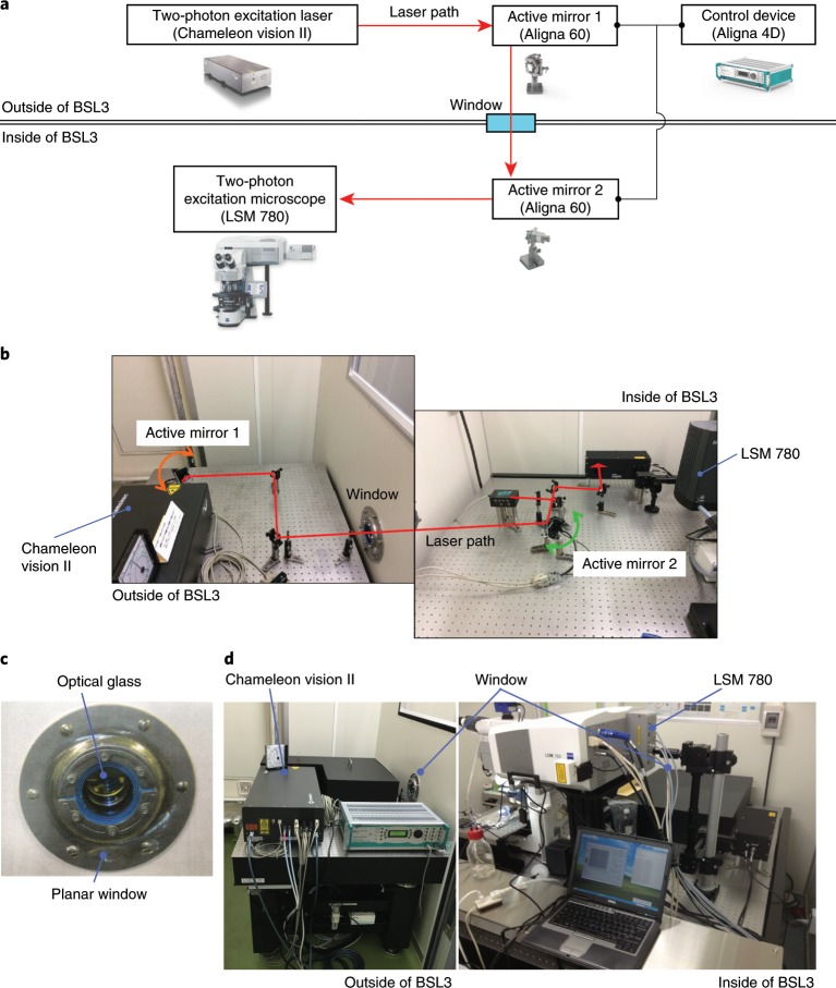
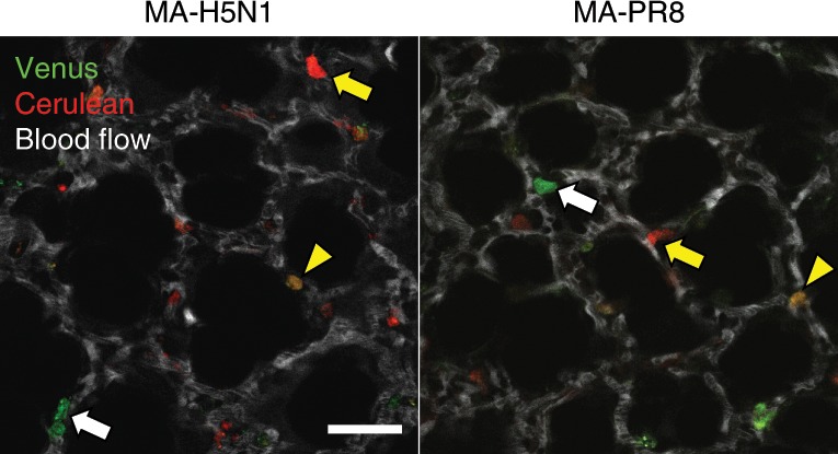
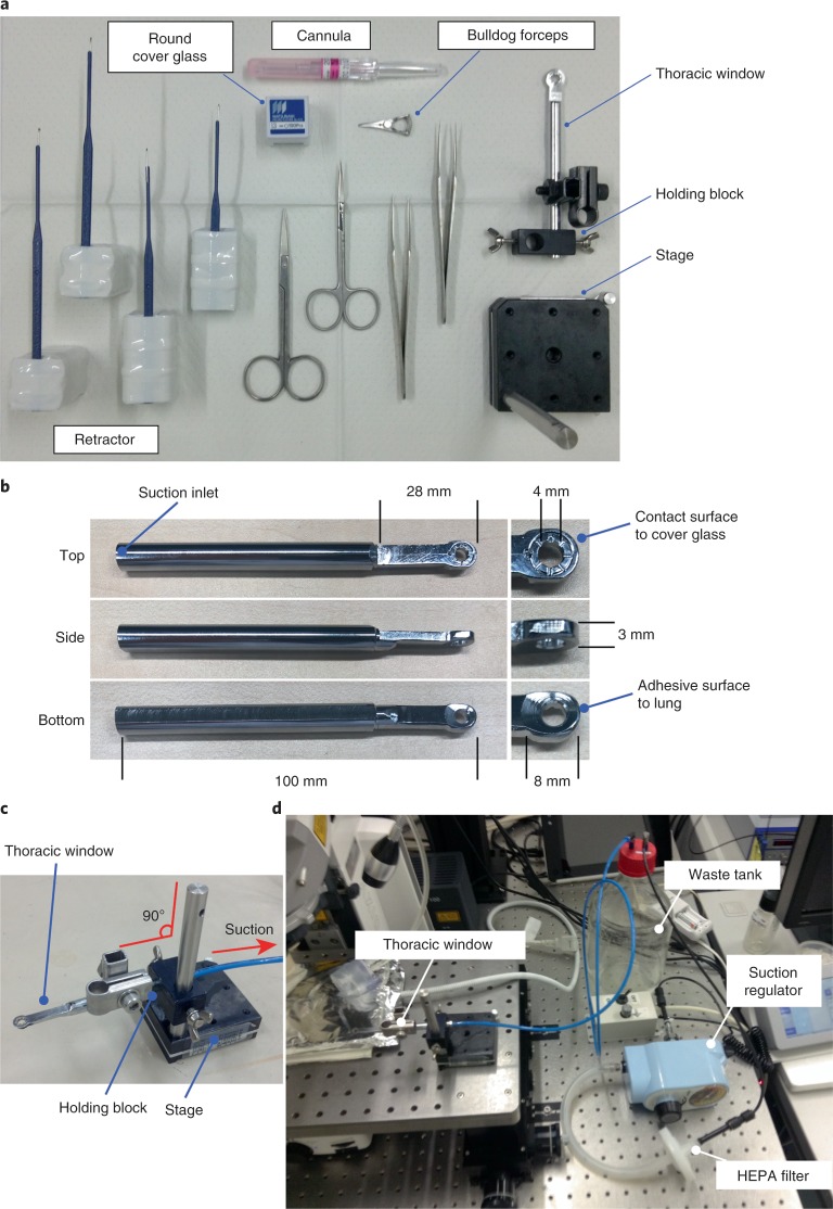
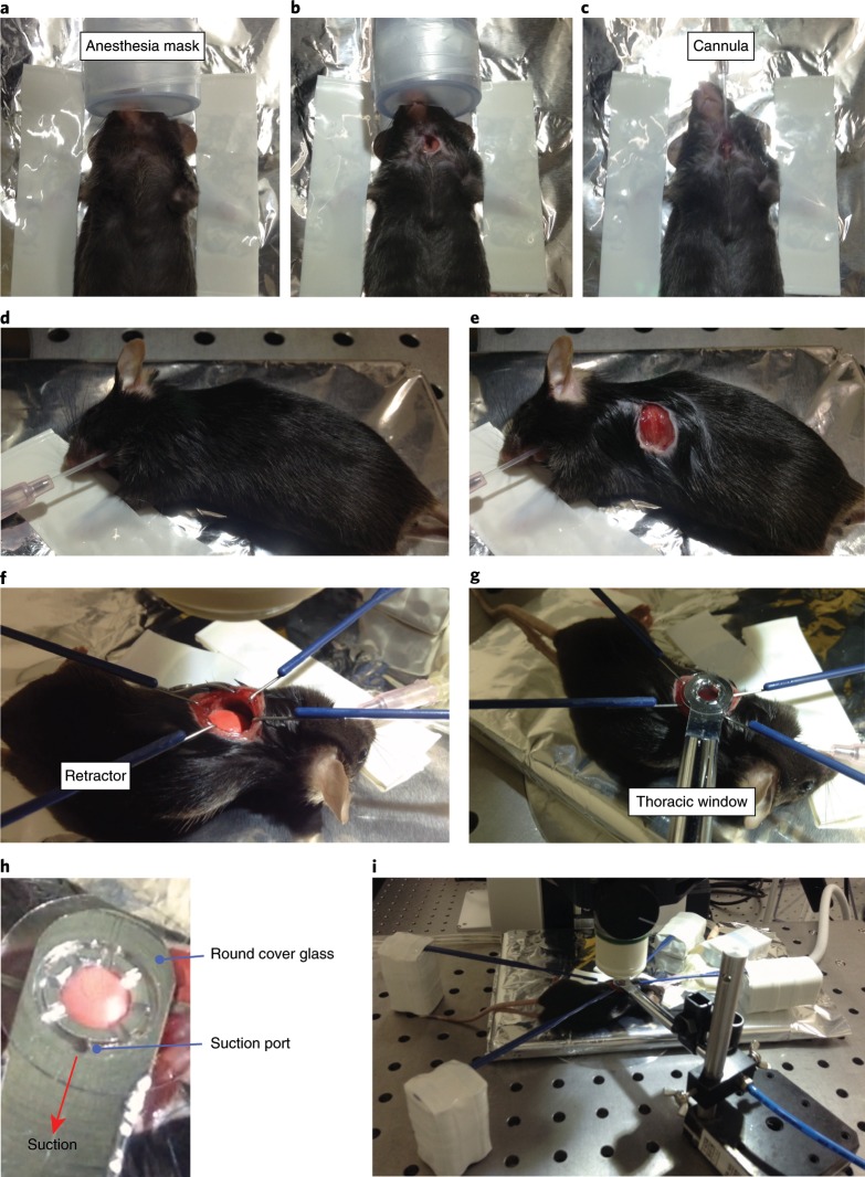
In vivo two-photon imaging is a valuable technique for studies of viral pathogenesis and host responses to infection in vivo. In this protocol, we describe a methodology for analyzing influenza virus-infected lung in vivo by two-photon imaging microscopy. We describe the surgical procedure, how to stabilize the lung, and an approach to analyzing the data. Further, we provide a database of fluorescent dyes, antibodies, and reporter mouse lines that can be used in combination with a reporter influenza virus (Color-flu) for multicolor analysis. Setup of this model typically takes ~30 min and enables the observation of influenza virus-infected lungs for >4 h during the acute phase of the inflammation and at least 1 h in the lethal phase. This imaging system, which we termed two-photon IMPRESS (imaging pathophysiology research system), is broadly applicable to analyses of other respiratory pathogens and reveals disease progression at the cellular level in vivo.

摘要

体内双光子成像是研究病毒发病机制和宿主对感染反应的一种有价值的技术。在本方案中,我们描述了一种通过双光子成像显微镜分析体内流感病毒感染肺的方法。我们描述了手术过程、如何稳定肺部,以及分析数据的方法。此外,我们提供了一个荧光染料、抗体和报告小鼠系的数据库,可与报告流感病毒(Color-flu)结合使用,进行多色分析。该模型的设置通常需要约 30 分钟,可在炎症的急性期观察感染流感病毒的肺超过 4 小时,在致死期至少 1 小时。我们将这种成像系统称为双光子 IMPRESS(成像病理生理学研究系统),它广泛适用于其他呼吸道病原体的分析,并在体内从细胞水平揭示疾病进展。

https://cdn.ncbi.nlm.nih.gov/pmc/blobs/1ea3/7086515/4348d99fcdd6/41596_2019_275_Fig6_HTML.jpg
https://cdn.ncbi.nlm.nih.gov/pmc/blobs/1ea3/7086515/8fcefaf092ae/41596_2019_275_Fig1_HTML.jpg
https://cdn.ncbi.nlm.nih.gov/pmc/blobs/1ea3/7086515/ac88c6509608/41596_2019_275_Fig2_HTML.jpg
https://cdn.ncbi.nlm.nih.gov/pmc/blobs/1ea3/7086515/0dc1262e4825/41596_2019_275_Fig3_HTML.jpg
https://cdn.ncbi.nlm.nih.gov/pmc/blobs/1ea3/7086515/8010b379b2a7/41596_2019_275_Fig4_HTML.jpg
https://cdn.ncbi.nlm.nih.gov/pmc/blobs/1ea3/7086515/aa0d81851d8c/41596_2019_275_Fig5_HTML.jpg
https://cdn.ncbi.nlm.nih.gov/pmc/blobs/1ea3/7086515/4348d99fcdd6/41596_2019_275_Fig6_HTML.jpg

相似文献

1
Multicolor two-photon imaging of in vivo cellular pathophysiology upon influenza virus infection using the two-photon IMPRESS.利用双光子 IMPRESS 技术对活体细胞内流感病毒感染后的病理生理学进行双色双光子成像。
Nat Protoc. 2020 Mar;15(3):1041-1065. doi: 10.1038/s41596-019-0275-y. Epub 2020 Jan 29.
2
Optimisations and Challenges Involved in the Creation of Various Bioluminescent and Fluorescent Influenza A Virus Strains for In Vitro and In Vivo Applications.用于体外和体内应用的各种生物发光和荧光甲型流感病毒株创建过程中涉及的优化与挑战。
PLoS One. 2015 Aug 4;10(8):e0133888. doi: 10.1371/journal.pone.0133888. eCollection 2015.
3
A Luciferase-fluorescent Reporter Influenza Virus for Live Imaging and Quantification of Viral Infection.一种用于病毒感染实时成像和定量分析的荧光素酶-荧光报告基因流感病毒。
J Vis Exp. 2019 Aug 14(150). doi: 10.3791/59890.
4
Directed Evolution of an Influenza Reporter Virus To Restore Replication and Virulence and Enhance Noninvasive Bioluminescence Imaging in Mice.定向进化流感报告病毒以恢复复制和毒力并增强小鼠的非侵入性生物发光成像。
J Virol. 2018 Jul 31;92(16). doi: 10.1128/JVI.00593-18. Print 2018 Aug 15.
5
RNA-Seq Analysis of Influenza A Virus-Induced Transcriptional Changes in Mice Lung and Its Possible Implications for the Virus Pathogenicity in Mice.RNA-Seq 分析甲型流感病毒诱导的小鼠肺部转录变化及其对小鼠病毒致病性的可能影响。
Viruses. 2021 Oct 8;13(10):2031. doi: 10.3390/v13102031.
6
Replication-Competent ΔNS1 Influenza A Viruses Expressing Reporter Genes.复制型 ΔNS1 流感 A 病毒表达报告基因。
Viruses. 2021 Apr 17;13(4):698. doi: 10.3390/v13040698.
7
Visualizing influenza virus infection in living mice.在活鼠体内可视化流感病毒感染。
Nat Commun. 2013;4:2369. doi: 10.1038/ncomms3369.
8
Multi-spectral fluorescent reporter influenza viruses (Color-flu) as powerful tools for in vivo studies.多光谱荧光报告流感病毒(彩色流感病毒)作为体内研究的强大工具。
Nat Commun. 2015 Mar 25;6:6600. doi: 10.1038/ncomms7600.
9
A pharmacologically immunosuppressed mouse model for assessing influenza B virus pathogenicity and oseltamivir treatment.一种评估乙型流感病毒致病性和奥司他韦治疗效果的药理学免疫抑制小鼠模型。
Antiviral Res. 2017 Dec;148:20-31. doi: 10.1016/j.antiviral.2017.10.021. Epub 2017 Oct 31.
10
Transcriptome Analysis of Infected and Bystander Type 2 Alveolar Epithelial Cells during Influenza A Virus Infection Reveals Wnt Pathway Downregulation.转录组分析感染和旁观者 2 型肺泡上皮细胞在甲型流感病毒感染时揭示 Wnt 通路下调。
J Virol. 2018 Oct 12;92(21). doi: 10.1128/JVI.01325-18. Print 2018 Nov 1.

引用本文的文献

1
Finding and filling the knowledge gaps in mechanisms of T cell-mediated TB immunity to inform vaccine design.发现并填补T细胞介导的结核病免疫机制中的知识空白,为疫苗设计提供依据。
Nat Rev Immunol. 2025 Jun 13. doi: 10.1038/s41577-025-01192-z.
2
Molecular probes for in vivo optical imaging of immune cells.用于免疫细胞体内光学成像的分子探针。
Nat Biomed Eng. 2025 May;9(5):618-637. doi: 10.1038/s41551-024-01275-7. Epub 2025 Feb 21.
3
TWINKLE: An open-source two-photon microscope for teaching and research.TWINKLE:一款用于教学与研究的开源双光子显微镜。

本文引用的文献

1
Pulmonary environmental cues drive group 2 innate lymphoid cell dynamics in mice and humans.肺部环境线索驱动小鼠和人类 2 型固有淋巴细胞的动态变化。
Sci Immunol. 2019 Jun 7;4(36). doi: 10.1126/sciimmunol.aav7638.
2
Intravital imaging allows real-time characterization of tissue resident eosinophils.活体成像可实时描绘组织固有嗜酸性粒细胞。
Commun Biol. 2019 May 13;2:181. doi: 10.1038/s42003-019-0425-3. eCollection 2019.
3
Two distinct interstitial macrophage populations coexist across tissues in specific subtissular niches.两种不同的组织间巨噬细胞群体存在于特定的组织亚区隔龛位中。
PLoS One. 2025 Feb 13;20(2):e0318924. doi: 10.1371/journal.pone.0318924. eCollection 2025.
4
Neutrophil adhesion to vessel walls impairs pulmonary circulation in COVID-19 pathology.在新冠病毒疾病病理学中,中性粒细胞与血管壁的黏附会损害肺循环。
Nat Commun. 2025 Jan 13;16(1):455. doi: 10.1038/s41467-024-55272-0.
5
LungVis 1.0: an automatic AI-powered 3D imaging ecosystem unveils spatial profiling of nanoparticle delivery and acinar migration of lung macrophages.LungVis 1.0:一个自动的人工智能驱动的 3D 成像生态系统,揭示了纳米颗粒传递的空间分布和肺巨噬细胞的肺泡迁移。
Nat Commun. 2024 Nov 27;15(1):10138. doi: 10.1038/s41467-024-54267-1.
6
TWINKLE: An open-source two-photon microscope for teaching and research.TWINKLE:一款用于教学与研究的开源双光子显微镜。
bioRxiv. 2025 Jan 12:2024.09.23.612766. doi: 10.1101/2024.09.23.612766.
7
Multiphoton fluorescence microscopy for in vivo imaging.多光子荧光显微镜用于活体成像。
Cell. 2024 Aug 22;187(17):4458-4487. doi: 10.1016/j.cell.2024.07.036.
8
Dynamic landscapes and protective immunity coordinated by influenza-specific lung-resident memory CD8 T cells revealed by intravital imaging.通过活体成像揭示流感特异性肺驻留记忆 CD8 T 细胞协调的动态景观和保护性免疫。
Immunity. 2024 Aug 13;57(8):1878-1892.e5. doi: 10.1016/j.immuni.2024.06.016. Epub 2024 Jul 22.
9
Development of a Mouse-Adapted Reporter SARS-CoV-2 as a Tool for Two-Photon In Vivo Imaging.开发一种适应小鼠的报告基因 SARS-CoV-2 作为双光子体内成像工具。
Viruses. 2024 Mar 29;16(4):537. doi: 10.3390/v16040537.
10
NLRP3 inflammasome activation in sensory neurons promotes chronic inflammatory and osteoarthritis pain.感觉神经元中的NLRP3炎性小体激活会促进慢性炎症和骨关节炎疼痛。
Immunother Adv. 2023 Oct 24;3(1):ltad022. doi: 10.1093/immadv/ltad022. eCollection 2023.
Science. 2019 Mar 15;363(6432). doi: 10.1126/science.aau0964.
4
Neutrophils disturb pulmonary microcirculation in sepsis-induced acute lung injury.中性粒细胞在脓毒症诱导的急性肺损伤中扰乱肺部微循环。
Eur Respir J. 2019 Mar 28;53(3). doi: 10.1183/13993003.00786-2018. Print 2019 Mar.
5
Live imaging of the pulmonary immune environment.肺部免疫环境的活体成像。
Cell Immunol. 2020 Apr;350:103862. doi: 10.1016/j.cellimm.2018.09.007. Epub 2018 Oct 9.
6
Imaging Cell Interaction in Tracheal Mucosa During Influenza Virus Infection Using Two-photon Intravital Microscopy.利用双光子活体显微镜成像技术观察流感病毒感染期间气管黏膜中的细胞相互作用
J Vis Exp. 2018 Aug 17(138):58355. doi: 10.3791/58355.
7
α-Toxin Induces Platelet Aggregation and Liver Injury during Staphylococcus aureus Sepsis.α-毒素在金黄色葡萄球菌脓毒症中诱导血小板聚集和肝损伤。
Cell Host Microbe. 2018 Aug 8;24(2):271-284.e3. doi: 10.1016/j.chom.2018.06.017. Epub 2018 Jul 19.
8
Removing physiological motion from intravital and clinical functional imaging data.从活体和临床功能成像数据中去除生理运动。
Elife. 2018 Jul 9;7:e35800. doi: 10.7554/eLife.35800.
9
In vivo imaging of the pathophysiological changes and neutrophil dynamics in influenza virus-infected mouse lungs.体内成像研究流感病毒感染小鼠肺部的病理生理变化和中性粒细胞动力学。
Proc Natl Acad Sci U S A. 2018 Jul 10;115(28):E6622-E6629. doi: 10.1073/pnas.1806265115. Epub 2018 Jun 25.
10
Intravital imaging of a pulmonary endothelial surface layer in a murine sepsis model.小鼠脓毒症模型中肺内皮表面层的活体成像
Biomed Opt Express. 2018 Apr 26;9(5):2383-2393. doi: 10.1364/BOE.9.002383. eCollection 2018 May 1.