Eye Center, The Second Hospital of Jilin University, Changchun, China.
Jilin Provincial Key Laboratory on Molecular and Chemical Genetic, The Second Hospital of Jilin University, Changchun, China.
J Cell Mol Med. 2020 Mar;24(6):3346-3358. doi: 10.1111/jcmm.15008. Epub 2020 Jan 30.
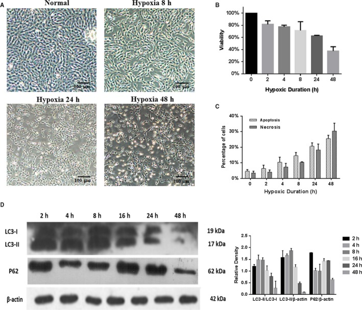
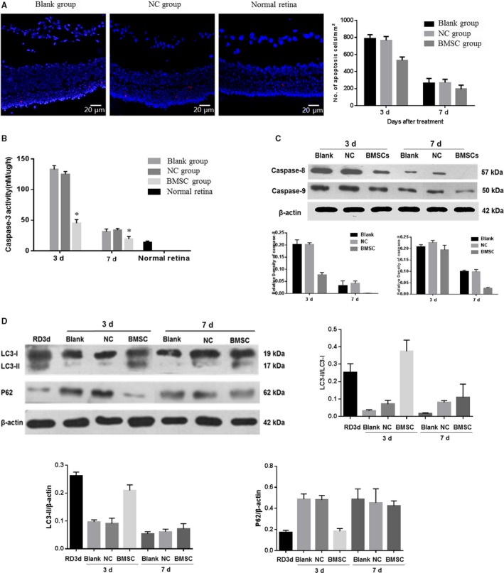
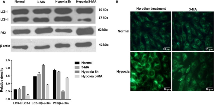
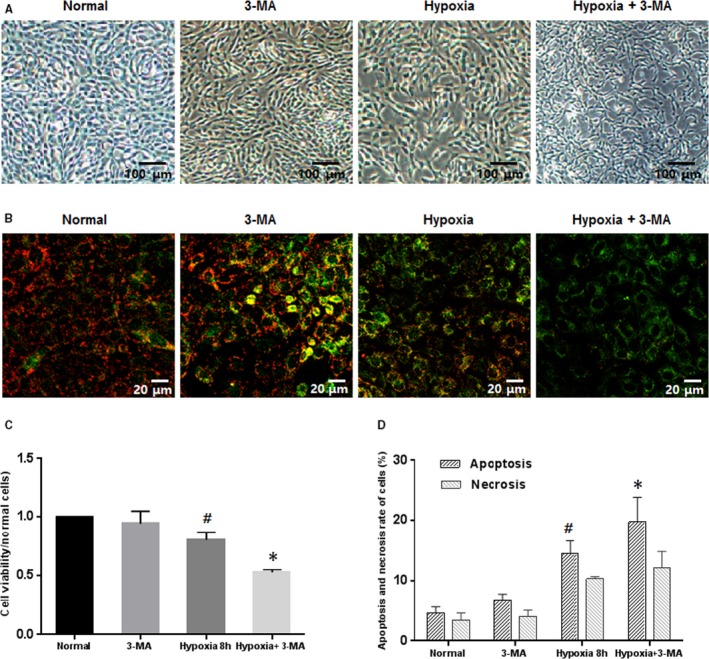
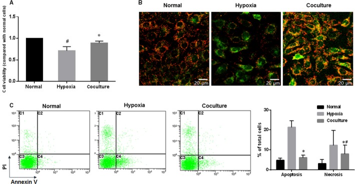
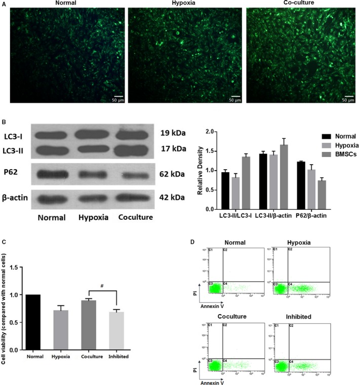
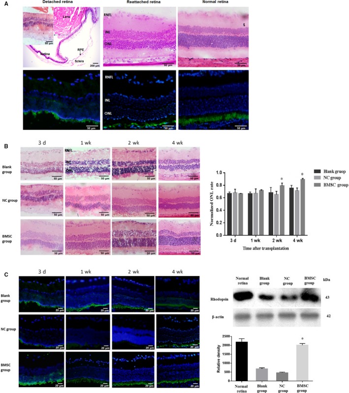
Our study aimed to evaluate the protective role and mechanisms of bone marrow mesenchymal stem cells (BMSCs) in hypoxic photoreceptors and experimental retinal detachment. The cellular morphology, viability, apoptosis and autophagy of hypoxic 661w cells and cells cocultured with BMSCs were analysed. In retinal detachment model, BMSCs were intraocularly transplanted, and then, the retinal morphology, outer nuclear layer (ONL) thickness and rhodopsin expression were studied as well as apoptosis and autophagy of the retinal cells. The hypoxia-induced apoptosis of 661w cells obviously increased together with autophagy levels increasing and peaking at 8 hours after hypoxia. Upon coculturing with BMSCs, hypoxic 661w cells had a better morphology and fewer apoptosis. After autophagy was inhibited, the apoptotic 661w cells under the hypoxia increased, and the cell viability was reduced, even in the presence of transplanted BMSCs. In retina-detached eyes transplanted with BMSCs, the retinal ONL thickness was closer to that of the normal retina. After transplantation, apoptosis decreased significantly and retinal autophagy was activated in the BMSC-treated retinas. Increased autophagy in the early stage could facilitate the survival of 661w cells under hypoxic stress. Coculturing with BMSCs protects 661w cells from hypoxic damage, possibly due to autophagy activation. In retinal detachment models, BMSC transplantation can significantly reduce photoreceptor cell death and preserve retinal structure. The capacity of BMSCs to reduce retinal cell apoptosis and to initiate autophagy shortly after transplantation may facilitate the survival of retinal cells in the low-oxygen and nutrition-restricted milieu after retinal detachment.
我们的研究旨在评估骨髓间充质干细胞(BMSCs)在缺氧光感受器和实验性视网膜脱离中的保护作用和机制。分析了缺氧 661w 细胞和与 BMSCs 共培养的细胞的细胞形态、活力、凋亡和自噬。在视网膜脱离模型中,BMSCs 被眼内移植,然后研究视网膜形态、外核层(ONL)厚度和视紫红质表达以及视网膜细胞的凋亡和自噬。661w 细胞的缺氧诱导凋亡明显增加,同时自噬水平增加,并在缺氧 8 小时后达到峰值。与 BMSCs 共培养后,缺氧 661w 细胞形态更好,凋亡更少。自噬被抑制后,缺氧下凋亡的 661w 细胞增加,细胞活力降低,即使存在移植的 BMSCs 也是如此。在移植 BMSCs 的视网膜脱离眼中,视网膜 ONL 厚度更接近正常视网膜。移植后,BMSC 处理的视网膜中凋亡明显减少,视网膜自噬被激活。早期增加自噬可以促进 661w 细胞在缺氧应激下的存活。与 BMSCs 共培养可以保护 661w 细胞免受缺氧损伤,这可能是由于自噬的激活。在视网膜脱离模型中,BMSC 移植可以显著减少光感受器细胞的死亡并保持视网膜结构。BMSC 移植后短时间内减少视网膜细胞凋亡和启动自噬的能力可能有助于视网膜脱离后低氧和营养受限环境中视网膜细胞的存活。