Barkur Surekha, Lukose Jijo, Chidangil Santhosh
Centre of Excellence for Biophotonics, Department of Atomic and Molecular Physics, Manipal Academy of Higher Education, Manipal, Karnataka 576104, India.
ACS Omega. 2020 Jan 10;5(3):1439-1447. doi: 10.1021/acsomega.9b02988. eCollection 2020 Jan 28.
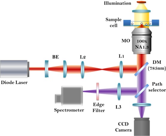
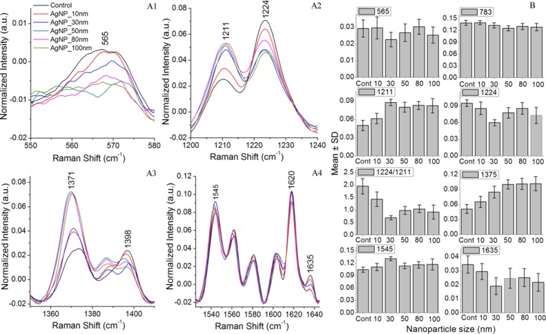
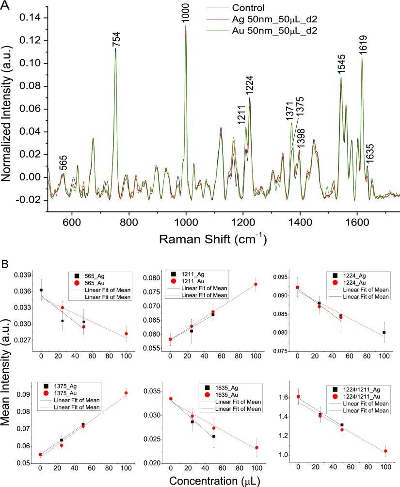
Advancements in the field of nanotechnology have resulted in the emergence of a large variety of engineered nanomaterials for innumerable applications. Despite the ubiquitous use of nanomaterials in daily life, concerns regarding the potential toxicity and safety of these materials have also been raised. There is a high demand for assessing the unwanted effects of both gold and silver nanoparticles, which is increasingly being used in biomedical applications. This paper deals with the study of stress due to silver and gold nanoparticles of varying size on red blood cells (RBCs) using Raman tweezers spectroscopy. RBCs were incubated with nanoparticles of size in the 10-100 nm range with the same concentrations, and micro-Raman spectra were recorded by optically trapping the nanoparticle-treated live RBCs. Spectral modifications implicating hemoglobin deoxygenation were observed in all nanoparticle-treated RBCs. One of the probable reason for the deoxygenation trend can be the adhesion of nanoparticles onto the cell surface causing imbalance in cell functioning. Moreover, the higher spectral variations observed for silver nanoparticles indicate that oxidative stress is involved in cell damage. These mechanisms lead to the modification in the hemoglobin structure because of changes in the pH of cytoplasm, which can be detected using Raman spectroscopy.
纳米技术领域的进步催生了大量用于无数应用的工程纳米材料。尽管纳米材料在日常生活中被广泛使用,但人们也对这些材料的潜在毒性和安全性表示担忧。对评估金纳米颗粒和银纳米颗粒的不良影响有很高的需求,这两种纳米颗粒在生物医学应用中越来越常用。本文利用拉曼镊子光谱研究了不同尺寸的银和金纳米颗粒对红细胞(RBC)产生的应力。将红细胞与尺寸在10 - 100纳米范围内、浓度相同的纳米颗粒一起孵育,通过光学捕获经纳米颗粒处理的活红细胞来记录显微拉曼光谱。在所有经纳米颗粒处理的红细胞中均观察到与血红蛋白脱氧相关的光谱变化。脱氧趋势的一个可能原因可能是纳米颗粒粘附在细胞表面导致细胞功能失衡。此外,银纳米颗粒观察到的更高光谱变化表明氧化应激参与了细胞损伤。这些机制由于细胞质pH值的变化导致血红蛋白结构发生改变,这可以通过拉曼光谱检测到。