Institut für Neuroimmunologie und Multiple Sklerose (INIMS), Universitätsklinikum Hamburg-Eppendorf (UKE), Martinistr. 52, Hamburg 20246, Germany.
Institut für Neuroimmunologie und Multiple Sklerose (INIMS), Universitätsklinikum Hamburg-Eppendorf (UKE), Martinistr. 52, Hamburg 20246, Germany.
Neuroimage Clin. 2020;25:102177. doi: 10.1016/j.nicl.2020.102177. Epub 2020 Jan 12.
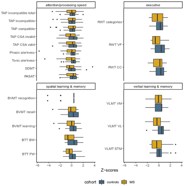
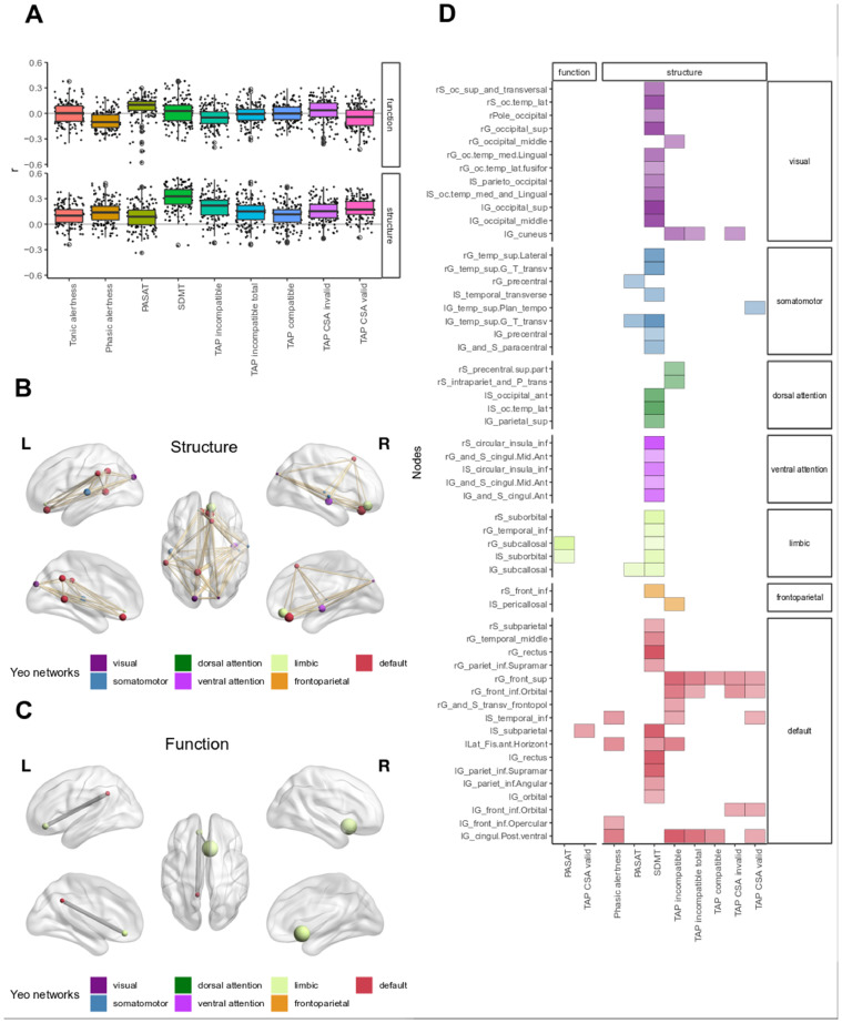
Multiple Sclerosis (MS) is the most common chronic inflammatory and neurodegenerative disease of the central nervous system (CNS), which can lead to severe cognitive impairment over time. Magnetic resonance imaging (MRI) is currently the best available biomarker to track MS pathophysiology in vivo and examine the link to clinical disability. However, conventional MRI metrics have limited sensitivity and specificity to detect direct associations between symptoms and their underlying CNS substrates. In this study, we aimed to investigate structural and resting state functional connectomes and subnetworks associated with neuropsychological (NP) performance using a graph theoretical approach. A comprehensive NP test battery was administered in a sample of patients with relapsing remitting MS (RRMS) and mild disability [n = 33, F/M = 20/13, age = 40.9 ± 9.7, median [Expanded Disability Status Scale] (EDSS) = 2, range =0-4] and compared to healthy controls (HC) [n = 29, F/M = 19/10, age = 41.0 ± 8.5] closely matched for age, sex, and level of education. The NP battery comprised the most relevant domains of cognitive dysfunction in MS including attention, processing speed, verbal and spatial learning and memory, and executive function. While standard MRI metrics showed good correlations with TAP Alertness test, disease duration and neurological exams, structural networks showed closer associations with 9-hole peg test and cognitive performances. Decreased graph strength was associated with two out of the 5 NP tests in the spatial learning and memory domain specified by BVMT [Sum 1-3] and BVMT [Recall], and with also SDMT which is one out of the 9 NP tests in the attention/processing speed domain, while no correlation was found between these scores and functional connectivity. Nodal strength was decreased in all subnetworks based on Yeo atlas in patients compared to HC; however, no difference was observed in nodal level of functional connectivity between the groups. The difference in structural and functional nodal connectivity between the groups was also observed in the relationship between structural and functional connectivity within the groups; the relationship between nodal degree and nodal strength was reversed in patients but positive in controls. On a nodal level, structural and functional networks (mainly the default mode network) were correlated with more than one cognitive domain rather than one specific network for each domain within patients. Interestingly, poorer cognitive performance was mostly correlated with increased functional connectivity but decreased structural connectivity in patients. Increased functional connectivity in the default mode network had both positive as well as negative associations with verbal and spatial learning and memory, possibly indicating adaptive and maladaptive mechanisms. In conclusion, our results suggest that cognitive performance, even in patients with RRMS and very mild disability, may reflect a loss of structural connectivity. In contrast, widespread increases in functional connectivity may be the result of maladaptive processes.
多发性硬化症 (MS) 是中枢神经系统 (CNS) 最常见的慢性炎症性和神经退行性疾病,随着时间的推移可导致严重的认知障碍。磁共振成像 (MRI) 目前是跟踪 MS 病理生理学和检查与临床残疾相关联的最佳生物标志物。然而,传统的 MRI 指标对检测症状与其潜在的中枢神经系统基质之间的直接关联的敏感性和特异性有限。在这项研究中,我们旨在使用图论方法研究与神经心理学 (NP) 表现相关的结构和静息状态功能连接组和子网。对一组复发性缓解型多发性硬化症 (RRMS) 且残疾程度较轻的患者 (n=33,F/M=20/13,年龄=40.9±9.7,中位数[扩展残疾状况量表] (EDSS)=2,范围为 0-4) 和健康对照组 (HC) [n=29,F/M=19/10,年龄=41.0±8.5] 进行了综合 NP 测试,这些对照组在年龄、性别和教育程度方面与患者密切匹配。NP 测试包括与 MS 相关的认知功能障碍的最相关领域,包括注意力、处理速度、言语和空间学习和记忆以及执行功能。虽然标准 MRI 指标与 TAP 警觉测试、疾病持续时间和神经检查具有良好的相关性,但结构网络与 9 孔钉测试和认知表现具有更密切的关联。在空间学习和记忆领域的 BVMT [总和 1-3] 和 BVMT [回忆] 中指定的 5 项 NP 测试中的 2 项以及注意力/处理速度领域的 9 项 NP 测试中的 1 项中,与认知表现相关的图形强度降低,而这些分数与功能连接之间没有相关性。与 HC 相比,患者的所有 Yeo 图谱子网中的节点强度都降低了;然而,两组之间的功能连接的节点水平没有差异。在组内结构和功能节点连接之间也观察到了组间结构和功能节点连接的差异;在患者中,节点度和节点强度之间的关系发生了逆转,但在对照组中则为正相关。在节点水平上,结构和功能网络 (主要是默认模式网络) 与多个认知领域相关,而不是与患者中每个领域的特定网络相关。有趣的是,在患者中,较差的认知表现与功能连接增加但结构连接减少有关。默认模式网络中功能连接的增加与言语和空间学习和记忆既有正相关也有负相关,这可能表明存在适应性和适应性不良的机制。总之,我们的结果表明,认知表现,即使在 RRMS 且残疾程度非常轻微的患者中,也可能反映出结构连接的丧失。相比之下,功能连接的广泛增加可能是适应不良过程的结果。