Bussi Simona, Coppo Alessandra, Celeste Roberto, Fanizzi Antonello, Fringuello Mingo Alberto, Ferraris Andrea, Botteron Catherine, Kirchin Miles A, Tedoldi Fabio, Maisano Federico
Bracco Imaging SpA, Bracco Research Centre, Via Ribes 5, 10010, Colleretto Giacosa, TO, Italy.
Bracco SpA, Via Caduti di Marcinelle 13, 20134, Milano, Italy.
Insights Imaging. 2020 Feb 4;11(1):11. doi: 10.1186/s13244-019-0824-5.
The purpose of this study was to compare Gd levels in rat tissues after cumulative exposure to four commercially available macrocyclic gadolinium-based contrast agents (GBCAs).
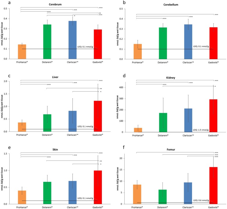
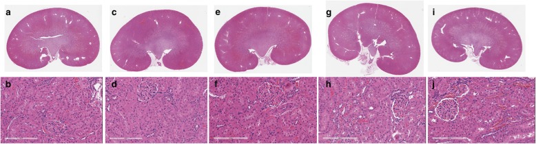
Sixty-five male Sprague-Dawley rats were randomized to four exposure groups (n = 15 per group) and one control group (n = 5). Animals in each exposure group received 20 GBCA administrations (four per week of ProHance®, Dotarem®, Clariscan™, or Gadovist® for 5 consecutive weeks) at a dose of 0.6 mmol/kg bodyweight. After 28-days' recovery, animals were sacrificed and tissues harvested for Gd determination by inductively coupled plasma-mass spectroscopy (ICP-MS). Histologic assessment of the kidney tissue was performed for all animals.
Significantly (p ≤ 0.005; all evaluations) lower Gd levels were noted with ProHance® than with Dotarem®, Clariscan™, or Gadovist® in all soft tissue organs: 0.144 ± 0.015 nmol/g vs. 0.342 ± 0.045, 0.377 ± 0.042, and 0.292 ± 0.047 nmol/g, respectively, for cerebrum; 0.151 ± 0.039 nmol/g vs. 0.315 ± 0.04, 0.345 ± 0.053, and 0.316 ± 0.040 nmol/g, respectively, for cerebellum; 0.361 ± 0.106 nmol/g vs. 0.685 ± 0.330, 0.823 ± 0.495, and 1.224 ± 0.664 nmol/g, respectively, for liver; 38.6 ± 25.0 nmol/g vs. 172 ± 134, 212 ± 121, and 294 ± 127 nmol/g, respectively, for kidney; and 0.400 ± 0.112 nmol/g vs. 0.660 ± 0.202, 0.688 ± 0.215, and 0.999 ± 0.442 nmol/g, respectively, for skin. No GBCA-induced macroscopic or microscopic findings were noted in the kidneys.
Less Gd is retained in the brain and body tissues of rats 28 days after the last exposure to ProHance® compared to other macrocyclic GBCAs, likely due to unique physico-chemical features that facilitate more rapid and efficient clearance.
本研究旨在比较大鼠在累积接触四种市售大环钆基造影剂(GBCAs)后组织中的钆含量。
65只雄性斯普拉格 - 道利大鼠被随机分为四个暴露组(每组n = 15)和一个对照组(n = 5)。每个暴露组的动物以0.6 mmol/kg体重的剂量接受20次GBCA给药(每周4次,连续5周分别给予普美显®、多它灵®、克沙醇™或钆布醇®)。在恢复28天后,处死动物并采集组织,通过电感耦合等离子体质谱法(ICP - MS)测定钆含量。对所有动物的肾组织进行组织学评估。
在所有软组织器官中,普美显®组的钆含量显著低于多它灵®、克沙醇™或钆布醇®组(p≤0.005;所有评估):大脑中分别为0.144±0.015 nmol/g 对比0.342±0.045、0.377±0.042和0.292±0.047 nmol/g;小脑分别为0.151±0.039 nmol/g对比0.315±0.04、0.345±0.053和0.316±0.040 nmol/g;肝脏分别为0.361±0.106 nmol/g对比0.685±0.330、0.823±0.495和1.224±0.664 nmol/g;肾脏分别为38.6±25.0 nmol/g对比172±134、212±121和294±127 nmol/g;皮肤分别为0.400±0.112 nmol/g对比0.660±0.202、0.688±0.215和0.999±0.442 nmol/g。在肾脏中未观察到GBCA引起的宏观或微观变化。
与其他大环GBCAs相比,大鼠在最后一次接触普美显® 28天后,大脑和身体组织中保留的钆较少,这可能是由于其独特的物理化学特性有助于更快速有效地清除。