Pai Vivek, Sitoh Yih Yian, Purohit Bela
Department of Neuroradiology, National Neuroscience Institute, 11 Jalan Tan Tock Seng, Singapore, 308433, Singapore.
Insights Imaging. 2020 Feb 10;11(1):20. doi: 10.1186/s13244-019-0829-0.
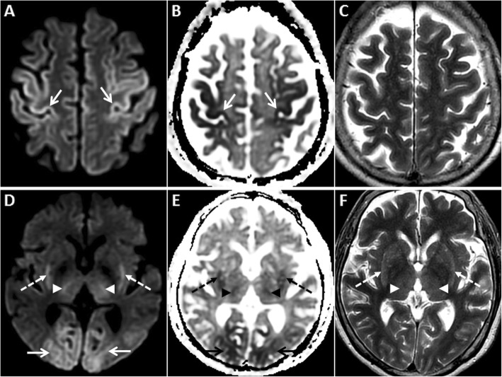
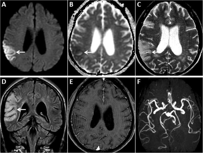
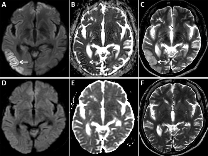
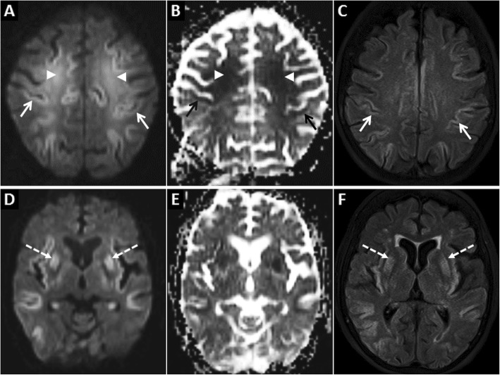
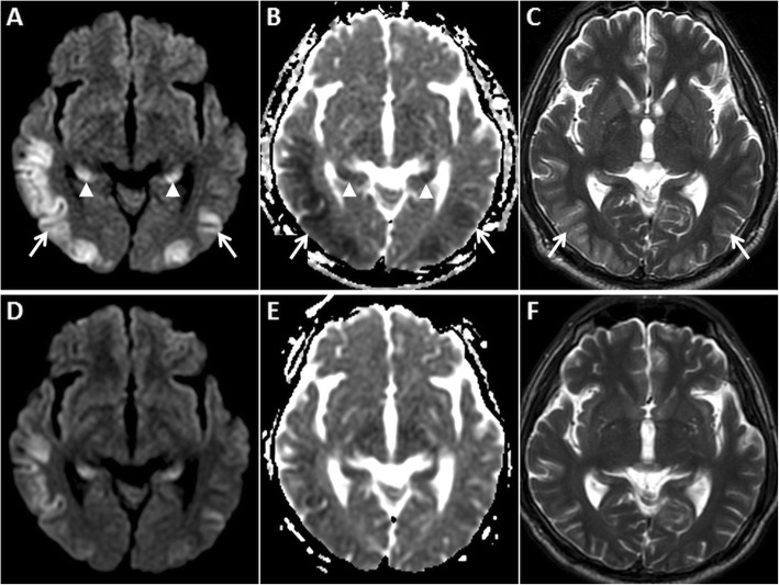
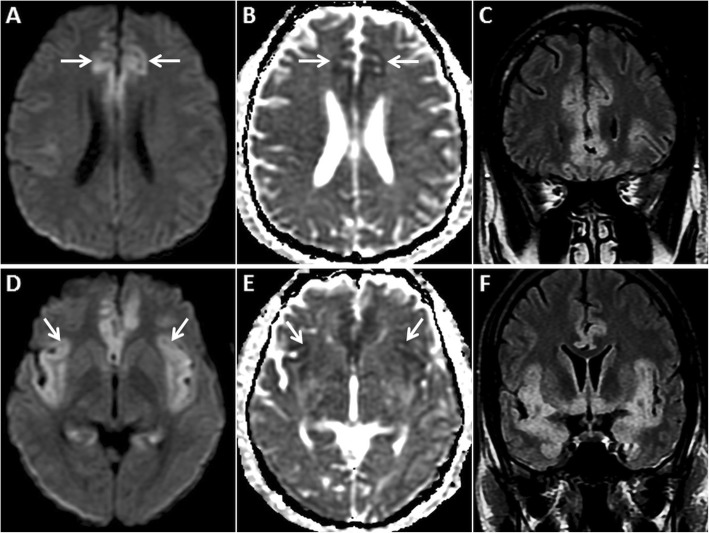
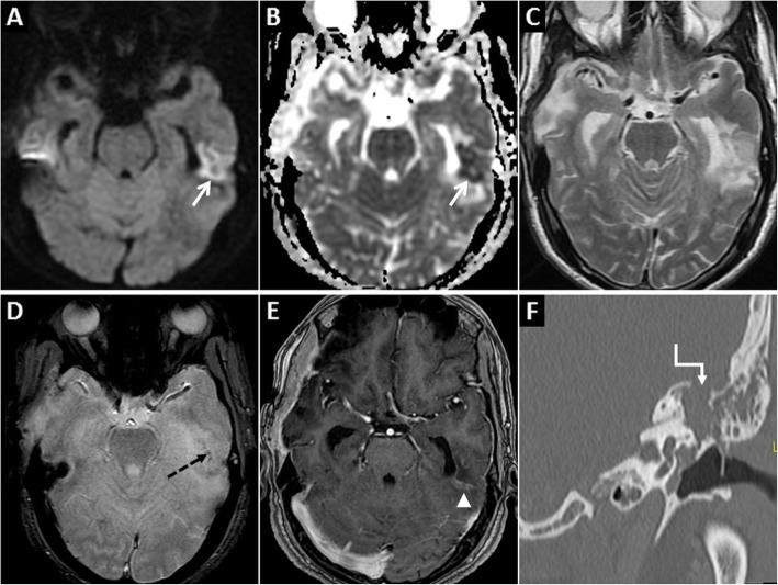
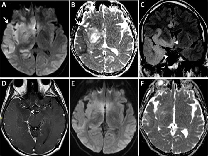
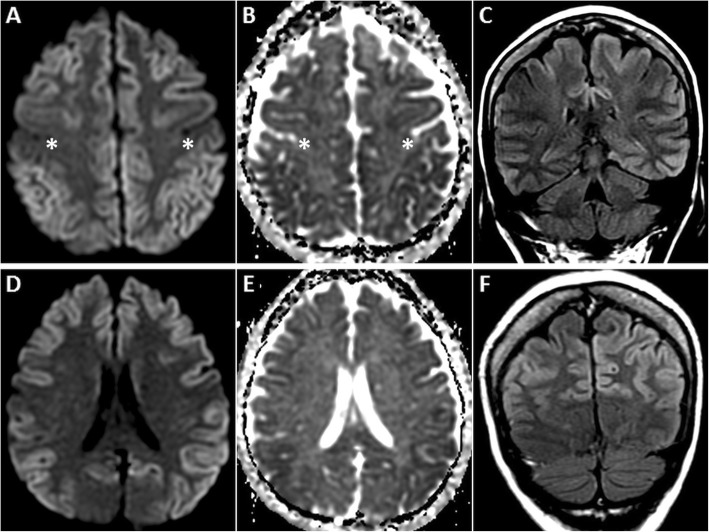
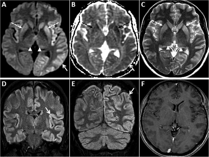
Gyriform restricted diffusion (GRD) refers to hyperintense signal involving the cerebral cortex on diffusion-weighted images (DWI) with corresponding hypointensity on apparent diffusion coefficient (ADC) images. These changes are commonly seen following a vascular occlusion, reflecting the limitation of water molecule movement across cell membranes (restricted diffusion) due to the failure of Na/K-ATPase pumps (cytotoxic oedema). However, GRD can occur in several other neurological conditions as well. A thorough understanding of these conditions and their anatomic predilection plays a critical role in identifying and differentiating them from vascular thrombo-occlusion, with impact towards appropriate clinical management. This review highlights the less commonly encountered, non-stroke causes of GRD in adults with case-based examples. A tabulated chart of the patterns of cortical and subcortical involvement associated with these aetiologies is provided for a quick, pattern-based reference for daily radiological reporting.
脑回状受限扩散(GRD)是指在扩散加权成像(DWI)上累及大脑皮质的高信号,在表观扩散系数(ADC)图像上相应为低信号。这些改变常见于血管闭塞后,反映由于钠钾ATP酶泵功能衰竭(细胞毒性水肿)导致水分子跨细胞膜移动受限(受限扩散)。然而,GRD也可出现在其他几种神经系统疾病中。全面了解这些疾病及其解剖学偏好对于将它们与血管血栓闭塞进行鉴别诊断至关重要,这对恰当的临床管理具有重要意义。本综述通过实例强调成人中较少见的非卒中原因导致的GRD。提供了一个表格,列出与这些病因相关的皮质和皮质下受累模式,以便在日常放射学报告中基于模式进行快速参考。