Artas Hakan, Okcesiz Izzet
Department of Radiology, Faculty of Medicine, Firat University, Elazığ, Turkey.
Arch Med Sci. 2019 Jan 11;16(1):58-65. doi: 10.5114/aoms.2018.81135. eCollection 2020.
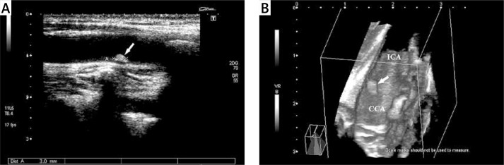
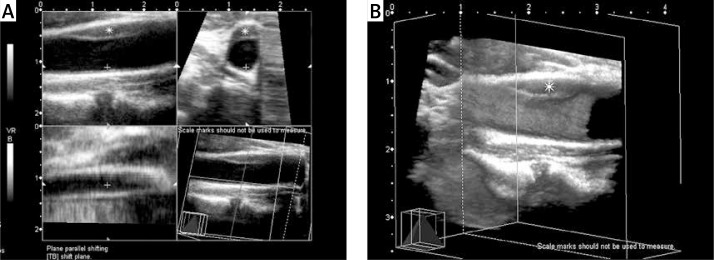
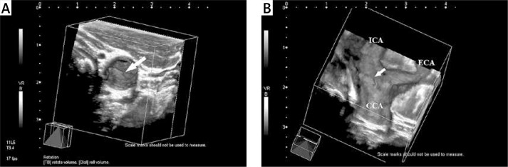
The aim of this study was to evaluate 3-dimensional (3D) ultrasonography (US) in determining the surface irregularity of carotid artery plaques.
This study included 50 patients (20 females and 30 males) aged between 56 and 82 years with plaques in the carotid artery which were detected during routine neck ultrasound. Simultaneously these cases were evaluated in terms of plaque echogenicities and surface characteristics with 2D and 3D US.
3D imaging was successfully performed in 45 of the 50 cases and the technical success rate was 90%. A single plaque was detected in 64.4% of the patients, with the remaining 35.6% having more than one plaque. The lengths of the plaques ranged from 2 to 12 mm (mean: 3.98 ±1.70 mm); the widths ranged from 1.8 to 3.2 mm (mean: 2.11 ±0.37 mm). No significant difference was found between 2D and 3D plaque echo-structures (observer 1, = 0.317; observer 2, = 0.276), but there were significant differences between 2D and 3D plaque surface irregularities (observer 1, = 0.002; observer 2, = 0.004). The inter-observer agreement on 2D and 3D plaque echo-structure and surface irregularity was very good (k coefficients were 0.89 and 0.83, respectively, for echo-structure, and 0.91 and 0.95, respectively, for surface irregularity).
The present study shows that 3D US examination is a valuable non-invasive method for investigation of surface irregularity of carotid artery plaques.
本研究的目的是评估三维(3D)超声检查(US)在确定颈动脉斑块表面不规则性方面的作用。
本研究纳入了50例年龄在56至82岁之间的患者(20例女性和30例男性),这些患者在常规颈部超声检查中发现有颈动脉斑块。同时,使用二维和三维超声对这些病例的斑块回声特性和表面特征进行评估。
50例患者中有45例成功进行了三维成像,技术成功率为90%。64.4%的患者检测到单个斑块,其余35.6%的患者有多个斑块。斑块长度范围为2至12毫米(平均:3.98±1.70毫米);宽度范围为1.8至3.2毫米(平均:2.11±0.37毫米)。二维和三维斑块回声结构之间未发现显著差异(观察者1,P = 0.317;观察者2,P = 0.276),但二维和三维斑块表面不规则性之间存在显著差异(观察者1,P = 0.002;观察者2,P = 0.004)。观察者之间在二维和三维斑块回声结构及表面不规则性方面的一致性非常好(回声结构的k系数分别为0.89和0.83,表面不规则性的k系数分别为0.91和0.95)。
本研究表明,三维超声检查是一种用于研究颈动脉斑块表面不规则性的有价值的非侵入性方法。