,.
.
Invest Ophthalmol Vis Sci. 2020 Feb 7;61(2):24. doi: 10.1167/iovs.61.2.24.
O-GlcNAcylation of cellular proteins contributes to the pathophysiology of diabetes and evidence supports a role for augmented O-GlcNAcylation in diabetic retinopathy. The aim of this study was to investigate the impact of the renin-angiotensin system on retinal protein O-GlcNAcylation.
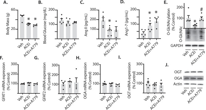
Mice fed a high-fat diet were treated chronically with the angiotensin-converting enzyme inhibitor captopril or captopril plus the angiotensin-(1-7) Mas receptor antagonist A779. Western blotting and quantitative polymerase chain reaction were used to analyze retinal homogenates. Similar analyses were performed on lysates from human MIO-M1 retinal Müller cell cultures exposed to media supplemented with angiotensin-(1-7). Culture conditions were manipulated to influence the hexosamine biosynthetic pathway and/or signaling downstream of the Mas receptor.
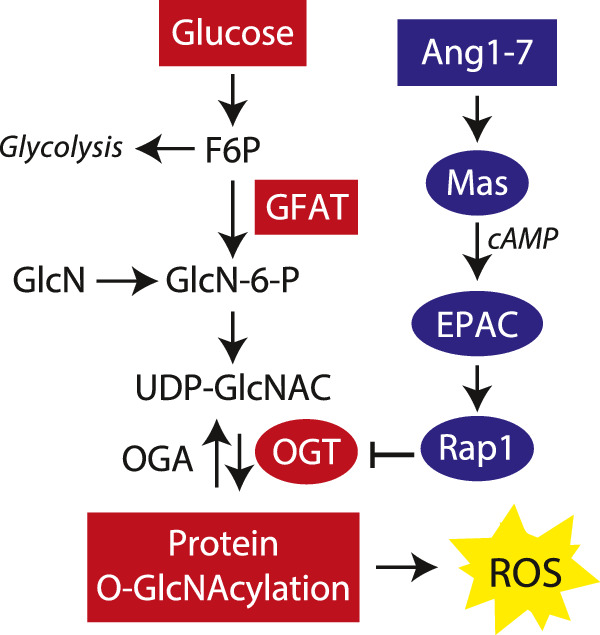
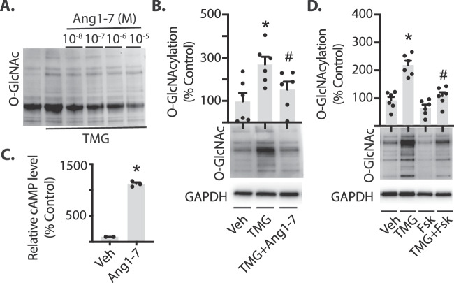
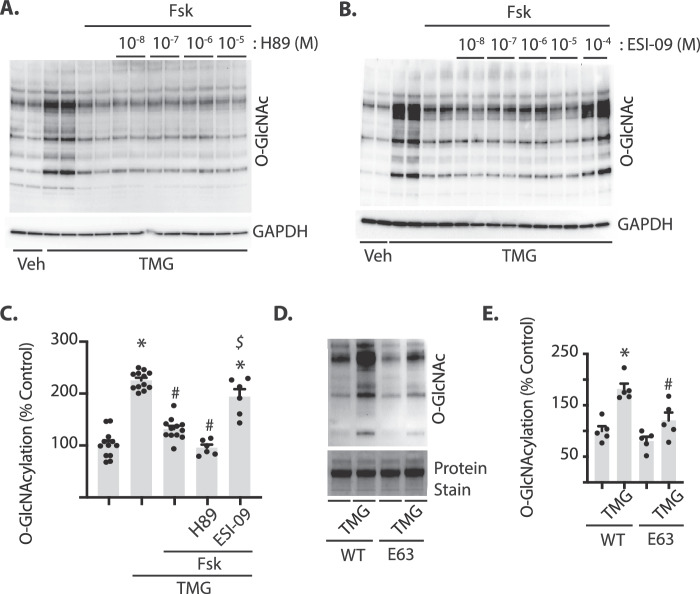
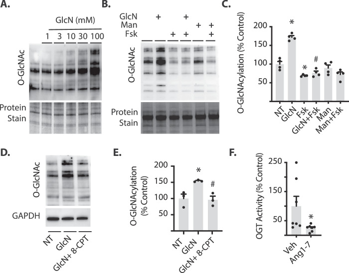
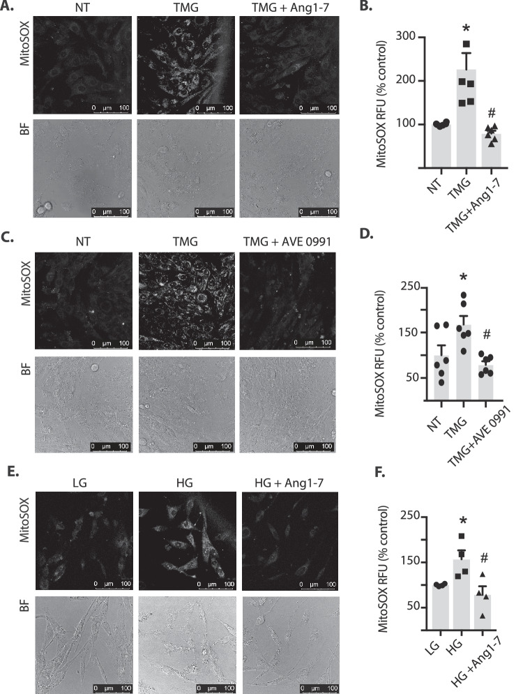
In the retina of mice fed a high-fat diet, captopril attenuated protein O-GlcNAcylation in a manner dependent on Mas receptor activation. In MIO-M1 cells, angiotensin-(1-7) or adenylate cyclase activation were sufficient to enhance cyclic AMP (cAMP) levels and inhibit O-GlcNAcylation. The repressive effect of cAMP on O-GlcNAcylation was dependent on exchange protein activated by cAMP (EPAC), but not protein kinase A, and was recapitulated by a constitutively active variant of the small GTPase Rap1. We provide evidence that cAMP and angiotensin-(1-7) act to suppress O-GlcNAcylation by inhibition of O-GlcNAc transferase (OGT) activity. In cells exposed to an O-GlcNAcase inhibitor or hyperglycemic culture conditions, mitochondrial superoxide levels were elevated; however, angiotensin-(1-7) signaling prevented the effect.
Angiotensin-(1-7) inhibits retinal protein O-GlcNAcylation via an EPAC/Rap1/OGT signaling axis.
细胞蛋白的 O-GlcNAc 化作用有助于糖尿病的病理生理学,有证据表明糖尿病性视网膜病变中 O-GlcNAc 化作用增强。本研究旨在探讨肾素-血管紧张素系统对视网膜蛋白 O-GlcNAc 化的影响。
用高脂肪饮食喂养小鼠,并用血管紧张素转换酶抑制剂卡托普利或卡托普利加血管紧张素-(1-7)Mas 受体拮抗剂 A779 进行慢性治疗。用 Western blot 和定量聚合酶链反应分析视网膜匀浆。对暴露于补充有血管紧张素-(1-7)的培养基的人 MIO-M1 视网膜 Müller 细胞培养物的裂解物进行类似分析。通过操纵培养条件来影响己糖胺生物合成途径和/或 Mas 受体下游的信号转导。
在高脂肪饮食喂养的小鼠视网膜中,卡托普利以依赖于 Mas 受体激活的方式减弱蛋白 O-GlcNAc 化。在 MIO-M1 细胞中,血管紧张素-(1-7)或腺苷酸环化酶激活足以增加环磷酸腺苷(cAMP)水平并抑制 O-GlcNAc 化。cAMP 对 O-GlcNAc 化的抑制作用依赖于 cAMP 激活的交换蛋白(EPAC),而不依赖于蛋白激酶 A,并且可以通过小 GTP 酶 Rap1 的组成性激活变体来再现。我们提供的证据表明,cAMP 和血管紧张素-(1-7)通过抑制 O-GlcNAc 转移酶(OGT)活性来抑制 O-GlcNAc 化。在暴露于 O-GlcNAcase 抑制剂或高糖培养条件的细胞中,线粒体超氧化物水平升高;然而,血管紧张素-(1-7)信号转导可防止这种作用。
血管紧张素-(1-7)通过 EPAC/Rap1/OGT 信号轴抑制视网膜蛋白 O-GlcNAc 化。