Department of Oral & Maxillofacial Surgery, University of Groningen, University Medical Center Groningen, Groningen, The Netherlands.
Oral Dis. 2021 Jan;27(1):21-26. doi: 10.1111/odi.13308. Epub 2020 Mar 13.
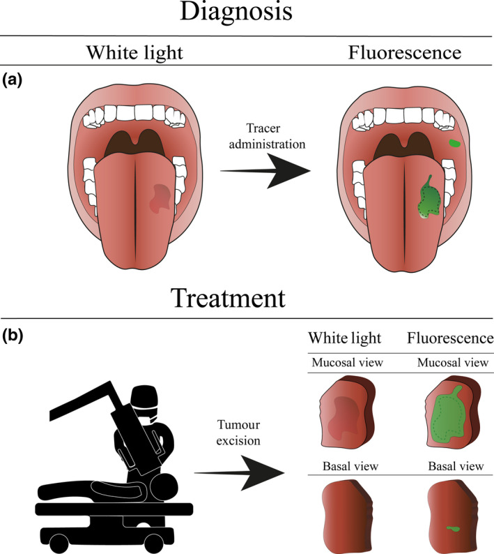
Early diagnosis and radical surgical excision of oral squamous cell carcinomas are essential for achieving optimal treatment outcomes. To date, diagnostic tools that rely on anatomical anomalies provide limited information and resolution in clinical practice. As a result, oral cancer is often detected in an advanced stage. Also, no reliable real-time intraoperative tools are readily available for the evaluation of surgical resection margins. Fluorescence imaging visualises biological processes that occur in early carcinogenesis and could, therefore, enable detection of small tumours in early stages. Furthermore, due to the high sensitivity and spatial resolution, fluorescence imaging could assist in resection margin assessment during surgery. In this review, we discuss several techniques that employ fluorescence for early diagnosis and surgical guidance in oral squamous cell carcinoma and present future perspectives on the potential of fluorescence imaging in oral cancer in the near future.
早期诊断和根治性手术切除口腔鳞状细胞癌对于获得最佳治疗效果至关重要。迄今为止,依靠解剖异常的诊断工具在临床实践中提供的信息和分辨率有限。因此,口腔癌通常在晚期才被发现。此外,目前还没有可靠的实时术中工具可用于评估手术切除边缘。荧光成像是对早期癌变过程中发生的生物学过程进行可视化,因此可以在早期阶段检测到小肿瘤。此外,由于具有高灵敏度和空间分辨率,荧光成像可以帮助评估手术中的切除边缘。在这篇综述中,我们讨论了几种利用荧光进行早期诊断和口腔鳞状细胞癌手术指导的技术,并对荧光成像在口腔癌中的近期应用前景进行了展望。