Garner Rikki M, Skariah Gemini, Hadjitheodorou Amalia, Belliveau Nathan M, Savinov Andrew, Footer Matthew J, Theriot Julie A
Biophysics Program, Stanford University School of Medicine, Stanford, CA.
Department of Biology, Howard Hughes Medical Institute, University of Washington, Seattle, WA.
Cytoskeleton (Hoboken). 2020 May;77(5-6):181-196. doi: 10.1002/cm.21603. Epub 2020 Feb 28.
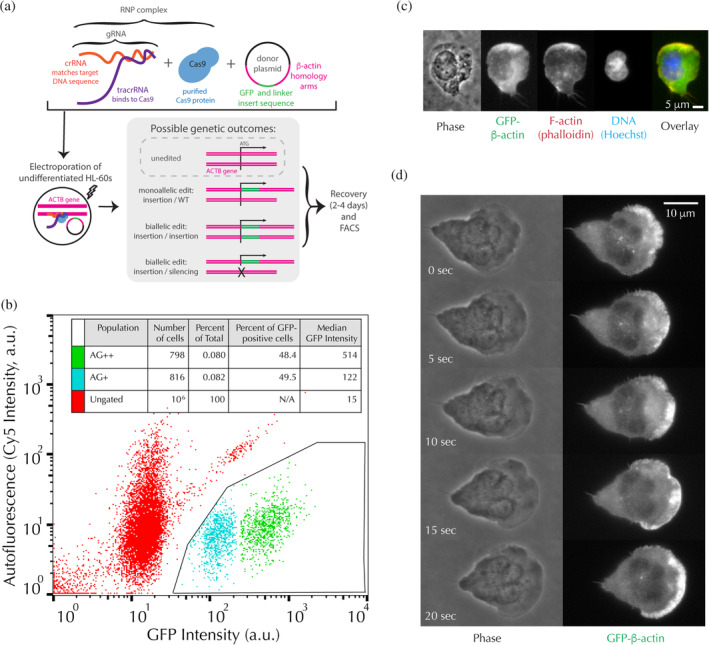
Observations of actin dynamics in living cells using fluorescence microscopy have been foundational in the exploration of the mechanisms underlying cell migration. We used CRISPR/Cas9 gene editing to generate neutrophil-like HL-60 cell lines expressing GFP-β-actin from the endogenous locus (ACTB). In light of many previous reports outlining functional deficiencies of labeled actin, we anticipated that HL-60 cells would only tolerate a monoallelic edit, as biallelic edited cells would produce no normal β-actin. Surprisingly, we recovered viable monoallelic GFP-β-actin cells as well as biallelic edited GFP-β-actin cells, in which one copy of the ACTB gene is silenced and the other contains the GFP tag. Furthermore, the edited cells migrate with similar speeds and persistence as unmodified cells in a variety of motility assays, and have nearly normal cell shapes. These results might partially be explained by our observation that GFP-β-actin incorporates into the F-actin network in biallelic edited cells at similar efficiencies as normal β-actin in unedited cells. Additionally, the edited cells significantly upregulate γ-actin, perhaps helping to compensate for the loss of normal β-actin. Interestingly, biallelic edited cells have only modest changes in global gene expression relative to the monoallelic line, as measured by RNA sequencing. While monoallelic edited cells downregulate expression of the tagged allele and are thus only weakly fluorescent, biallelic edited cells are quite bright and well-suited for live cell microscopy. The nondisruptive phenotype and direct interpretability of this fluorescent tagging approach make it a promising tool for studying actin dynamics in these rapidly migrating and highly phagocytic cells.
利用荧光显微镜对活细胞中的肌动蛋白动力学进行观察,一直是探索细胞迁移潜在机制的基础。我们使用CRISPR/Cas9基因编辑技术,从内源性位点(ACTB)生成表达GFP-β-肌动蛋白的中性粒细胞样HL-60细胞系。鉴于此前许多报道概述了标记肌动蛋白的功能缺陷,我们预计HL-60细胞只能耐受单等位基因编辑,因为双等位基因编辑的细胞不会产生正常的β-肌动蛋白。令人惊讶的是,我们获得了存活的单等位基因GFP-β-肌动蛋白细胞以及双等位基因编辑的GFP-β-肌动蛋白细胞,其中ACTB基因的一个拷贝被沉默,另一个含有GFP标签。此外,在各种运动分析中,编辑后的细胞与未修饰的细胞迁移速度和持续性相似,并且细胞形状几乎正常。这些结果可能部分可以通过我们的观察来解释,即GFP-β-肌动蛋白以与未编辑细胞中的正常β-肌动蛋白相似的效率掺入双等位基因编辑细胞的F-肌动蛋白网络中。此外,编辑后的细胞显著上调γ-肌动蛋白,这可能有助于补偿正常β-肌动蛋白的损失。有趣的是,通过RNA测序测量,双等位基因编辑细胞相对于单等位基因系在全局基因表达上只有适度变化。虽然单等位基因编辑细胞下调了标记等位基因的表达,因此荧光较弱,但双等位基因编辑细胞非常明亮,非常适合活细胞显微镜观察。这种荧光标记方法的无干扰表型和直接可解释性使其成为研究这些快速迁移和高度吞噬细胞中肌动蛋白动力学的有前途的工具。