Max Planck Institute for the Structure and Dynamics of Matter, CFEL, Luruper Chaussee 149, 22761, Hamburg, Germany.
Departments of Chemistry and Physics, University of Toronto, 80 St. George Street, Toronto, ON, M5S 3H6, Canada.
Nat Commun. 2020 Feb 21;11(1):996. doi: 10.1038/s41467-020-14793-0.
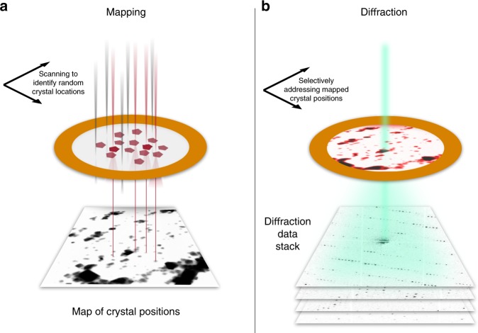
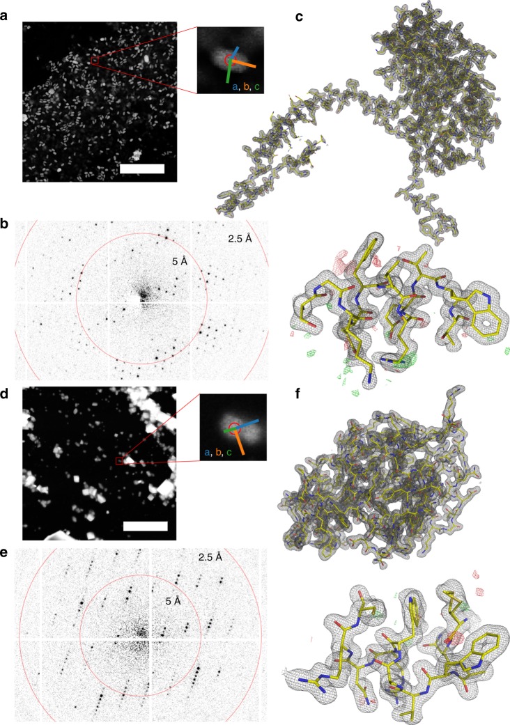
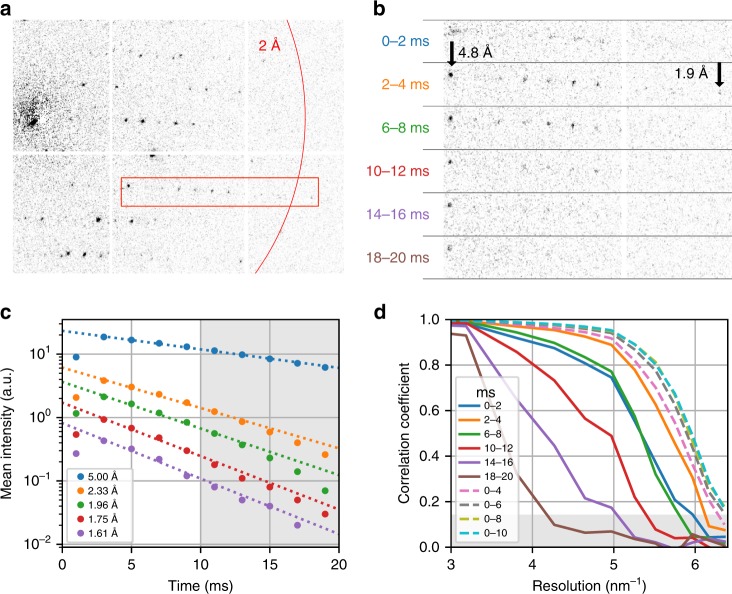
Serial X-ray crystallography at free-electron lasers allows to solve biomolecular structures from sub-micron-sized crystals. However, beam time at these facilities is scarce, and involved sample delivery techniques are required. On the other hand, rotation electron diffraction (MicroED) has shown great potential as an alternative means for protein nano-crystallography. Here, we present a method for serial electron diffraction of protein nanocrystals combining the benefits of both approaches. In a scanning transmission electron microscope, crystals randomly dispersed on a sample grid are automatically mapped, and a diffraction pattern at fixed orientation is recorded from each at a high acquisition rate. Dose fractionation ensures minimal radiation damage effects. We demonstrate the method by solving the structure of granulovirus occlusion bodies and lysozyme to resolutions of 1.55 Å and 1.80 Å, respectively. Our method promises to provide rapid structure determination for many classes of materials with minimal sample consumption, using readily available instrumentation.
利用自由电子激光进行连续 X 射线晶体学可以从小至亚微米尺寸的晶体中解析生物分子结构。然而,这些设施的光束时间稀缺,并且需要涉及样本传输技术。另一方面,旋转电子衍射(MicroED)已显示出作为蛋白质纳米晶体学的替代手段的巨大潜力。在这里,我们提出了一种将两种方法的优势相结合的用于蛋白质纳米晶体连续电子衍射的方法。在扫描透射电子显微镜中,随机分散在样品网格上的晶体自动进行映射,并以高采集率从每个晶体以固定取向记录衍射图案。剂量分割确保最小的辐射损伤效应。我们通过解析分别达到 1.55 Å 和 1.80 Å分辨率的颗粒病毒包埋体和溶菌酶的结构证明了该方法的可行性。我们的方法有望使用现成的仪器,以最小的样品消耗为许多类材料提供快速的结构测定。