Department of Respiratory and Critical Care Medicine, The First Affiliated Hospital, China Medical University, Shenyang 110001, China.
Mediators Inflamm. 2020 Feb 7;2020:4235909. doi: 10.1155/2020/4235909. eCollection 2020.
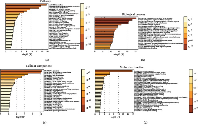
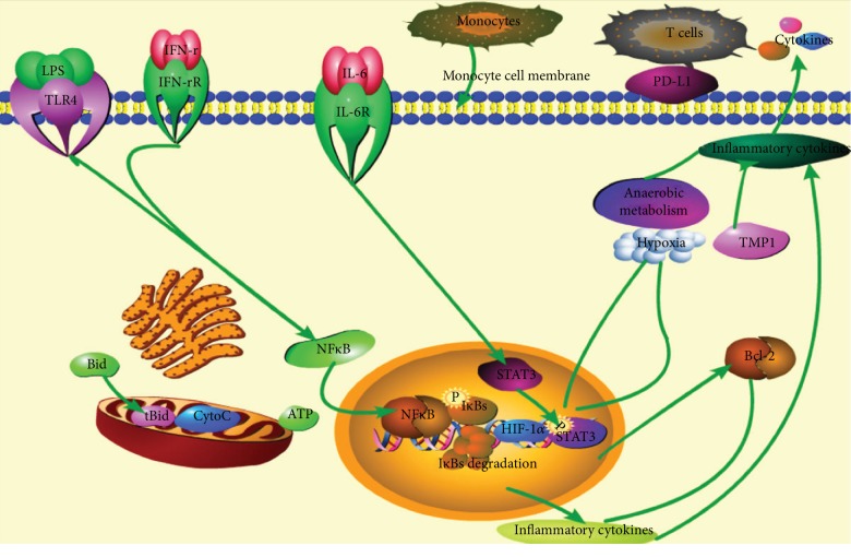
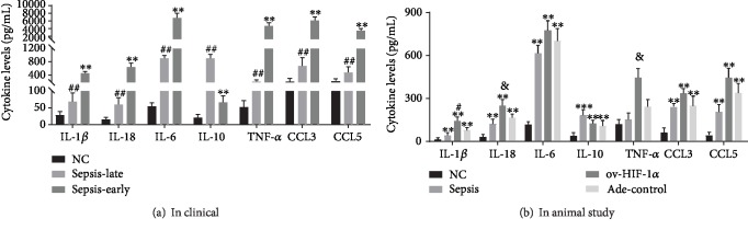
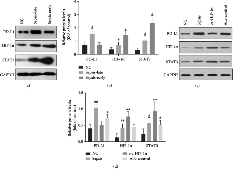
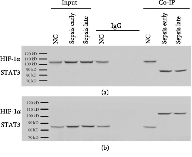
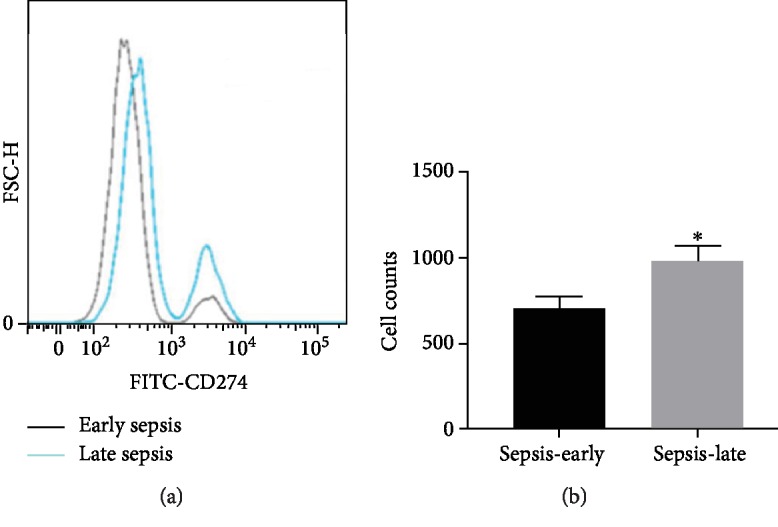
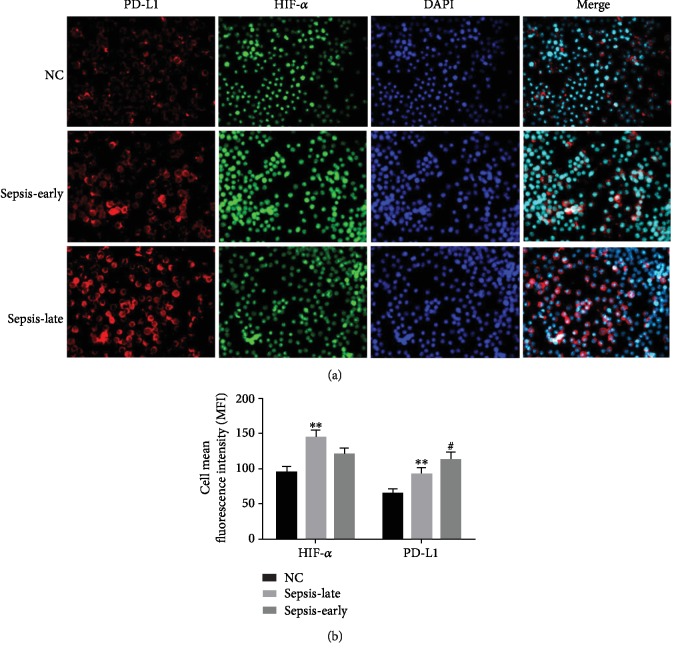
Severe pneumonia with sepsis is characterized by a dysregulated inflammatory response of endotoxin. In our study, we attempted to investigate the roles of the immune guardian cells (monocytes) in the immune-inflammatory response of severe pneumonia-induced sepsis. We performed analysis in the blood samples of human and animals with ELISA, western blot, flow cytometry (FCM) methods, etc. Results showed that the proinflammatory status shifted to hypoinflammatory phases during the sepsis process. In a clinical study, the levels of IL-1, IL-6, TNF-, etc., except for IL-10, were inhibited in the late phase of sepsis, while, in an animal study, the immune suppression status was attenuated with administration of the adenovirus Ade-HIF-1. Conversely, the amount of IL-10 was lower in the adenovirus Ade-HIF-1 group compared with the sepsis model group and the Ade-control group. Moreover, in the clinical study, the programmed cell death-ligand 1 (PD-L1) was overexpressed in monocytes in the late phase of sepsis, while the expression of proteins HIF-1 and STAT3 was decreased in the late phase of sepsis. However, in the animal study, we found that the HIF-1 factor facilitated the inflammatory response. The expression of the proteins HIF-1 and STAT3 was increased, and the PD-L1 protein was decreased with the adenovirus Ade-HIF-1 administration compared with the rats without Ade-HIF-1 injection and with the Ade-control injection. Additionally, the proteins HIF-1 and STAT3 were coregulated at transcriptional levels during the inflammatory responses of sepsis. Taken together, monocytes undergo reprogramming to generate immunosuppression through the HIF-1 signaling pathway in the late phase of sepsis.
严重肺炎伴败血症的特征是内毒素引起的炎症反应失调。在我们的研究中,我们试图研究免疫守护细胞(单核细胞)在严重肺炎引起的败血症的免疫炎症反应中的作用。我们通过 ELISA、western blot、流式细胞术(FCM)等方法分析了人类和动物的血液样本。结果表明,在败血症过程中,促炎状态向低炎症阶段转变。在临床研究中,除了 IL-10 之外,IL-1、IL-6、TNF- 等的水平在败血症晚期受到抑制,而在动物研究中,给予腺病毒 Ade-HIF-1 可减轻免疫抑制状态。相反,与败血症模型组和 Ade 对照组相比,腺病毒 Ade-HIF-1 组的 IL-10 水平较低。此外,在临床研究中,单核细胞在败血症晚期过度表达程序性细胞死亡配体 1(PD-L1),而在败血症晚期,HIF-1 和 STAT3 蛋白的表达减少。然而,在动物研究中,我们发现 HIF-1 因子促进了炎症反应。与未注射 Ade-HIF-1 的大鼠和注射 Ade 对照的大鼠相比,Ade-HIF-1 给药后 HIF-1 和 STAT3 蛋白的表达增加,PD-L1 蛋白减少。此外,在败血症的炎症反应中,HIF-1 和 STAT3 蛋白在转录水平上受到共同调控。总之,单核细胞在败血症晚期通过 HIF-1 信号通路发生重编程,产生免疫抑制。