Liu Jinjie, Liu Zanhua, Liu Guoliang, Gao Kai, Zhou Hengjie, Zhao Yongbo, Wang Hong, Zhang Lin, Liu Sibo
General Ward II, Affiliated Municipal Central Hospital of Dalian Medical University, Dalian, Liaoning 116000, P.R. China.
Department of Neurology, Nanjing Gaochun People's Hospital, Gaochun, Nanjing 210000, P.R. China.
Exp Ther Med. 2020 Mar;19(3):2103-2112. doi: 10.3892/etm.2020.8453. Epub 2020 Jan 15.
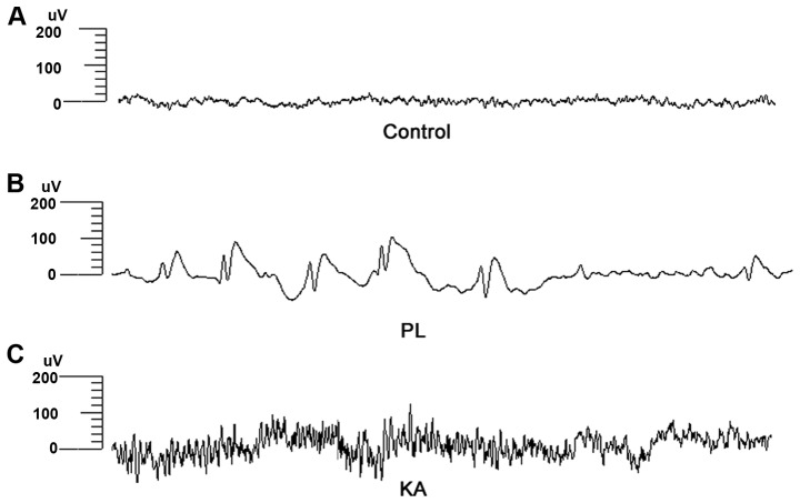
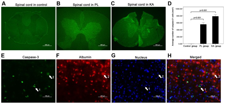
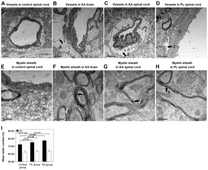
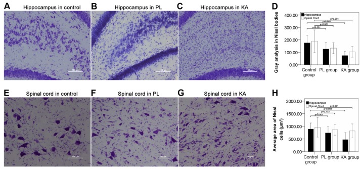
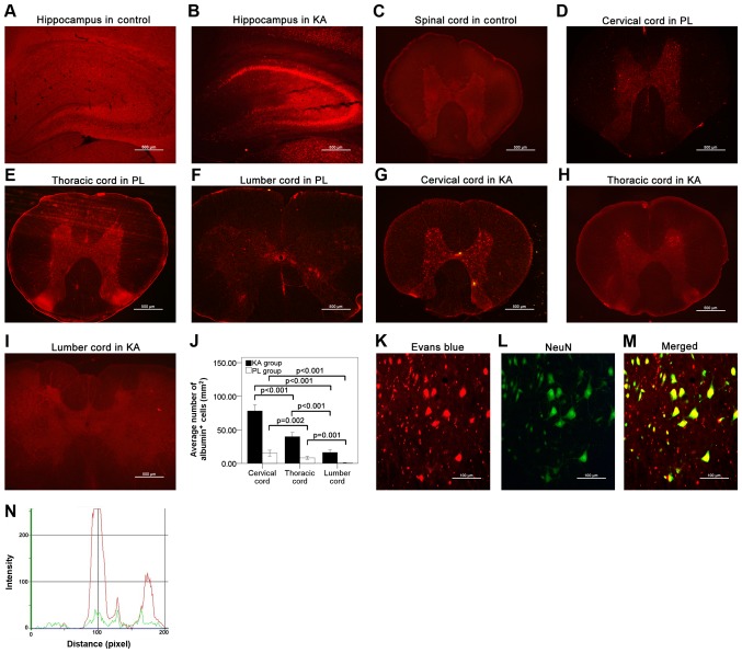
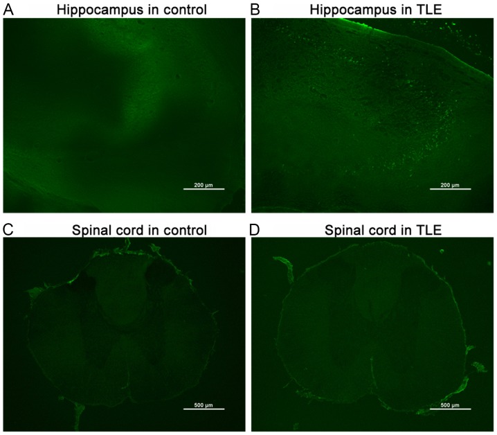
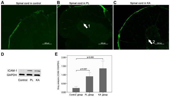
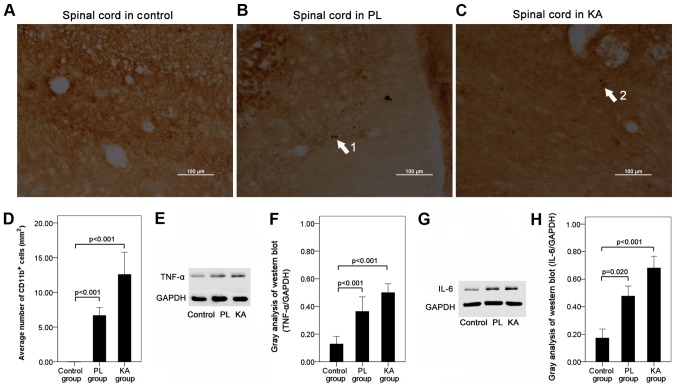
Numerous cases of spinal cord injury following seizure have been previously reported. However, whether spinal cord injury is a common occurrence after seizures and its underlying mechanisms remain unclear. The present study generated a Sprague-Dawley rat model of temporal lobe epilepsy (TLE), and Nissl staining and transmission electron microscopy were used to detect tissue damage. In addition, Evans blue staining was used to detect damage to the blood-brain barrier (BBB) and albumin extravasation. In addition, double-staining was used to detect the association between neurons and extravasated albumin. Furthermore, neuronal degeneration was assessed using Fluoro-Jade C staining, while fluorescence staining and western blotting were used to detect apoptosis and inflammation. In the present study, spinal cord injury was only observed in rats with grade IV-V seizures, whereas Nissl staining showed structural damage and decreased neuronal cell numbers in the brain and the spinal cord. The present study identified BBB damage and albumin extravasation in rats of the TLE groups. Double-staining for albumin and neurons showed a significant match of neurons positive for albumin. Fluoro-Jade C staining indicated neuronal degeneration in the brain, but not the spinal cord in the TLE rats. High levels of caspase-3 were also detected in the injured spinal cord. A small number of albumin neurons in the spinal cord presented caspase-3 signals in rats of the TLE groups. The expression levels of intercellular adhesion molecule 1, CD11b and inflammatory factors such as tumor necrosis factor-α and interleukin-6 were significantly elevated in the injured spinal cord. The present results suggested that spinal cord injury occurred in rats as a result of severe seizure attacks, and that BBB damage, albumin extravasation, inflammation and apoptosis contributed to the pathological changes observed during spinal cord injury.
先前已有许多癫痫发作后脊髓损伤的病例报道。然而,脊髓损伤在癫痫发作后是否常见及其潜在机制仍不清楚。本研究建立了颞叶癫痫(TLE)的Sprague-Dawley大鼠模型,采用尼氏染色和透射电子显微镜检测组织损伤。此外,采用伊文思蓝染色检测血脑屏障(BBB)损伤和白蛋白外渗。另外,采用双重染色检测神经元与外渗白蛋白之间的关联。此外,使用Fluoro-Jade C染色评估神经元变性,同时使用荧光染色和蛋白质免疫印迹法检测细胞凋亡和炎症。在本研究中,仅在IV-V级癫痫发作的大鼠中观察到脊髓损伤,而尼氏染色显示大脑和脊髓存在结构损伤且神经元细胞数量减少。本研究确定了TLE组大鼠存在BBB损伤和白蛋白外渗。白蛋白与神经元的双重染色显示白蛋白阳性的神经元有显著匹配。Fluoro-Jade C染色表明TLE大鼠大脑中的神经元发生变性,但脊髓未发生变性。在损伤的脊髓中还检测到高水平的半胱天冬酶-3。TLE组大鼠脊髓中的少量白蛋白神经元呈现半胱天冬酶-3信号。损伤脊髓中细胞间黏附分子1、CD11b以及肿瘤坏死因子-α和白细胞介素-6等炎性因子的表达水平显著升高。本研究结果表明,严重的癫痫发作导致大鼠发生脊髓损伤,并且BBB损伤、白蛋白外渗、炎症和细胞凋亡促成了脊髓损伤过程中观察到的病理变化。