Britton Chance Center for Biomedical Photonics, Wuhan National Laboratory for Optoelectronics-Huazhong University of Science and Technology, 430074, Wuhan, Hubei, China.
MoE Key Laboratory for Biomedical Photonics, Collaborative Innovation Center for Biomedical Engineering, School of Engineering Sciences, Huazhong University of Science and Technology, 430074, Wuhan, Hubei, China.
Nat Commun. 2020 Feb 28;11(1):1110. doi: 10.1038/s41467-020-14906-9.
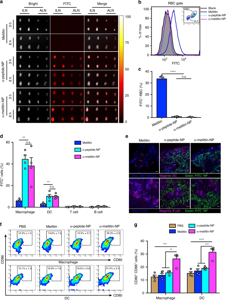
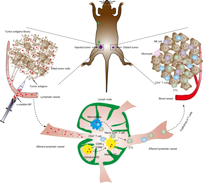
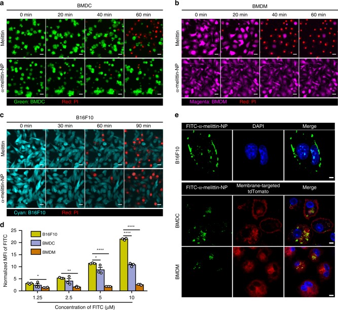
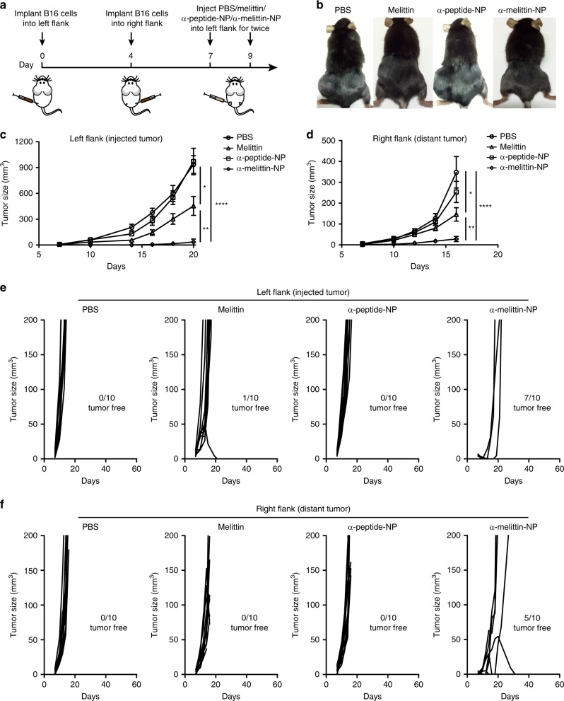
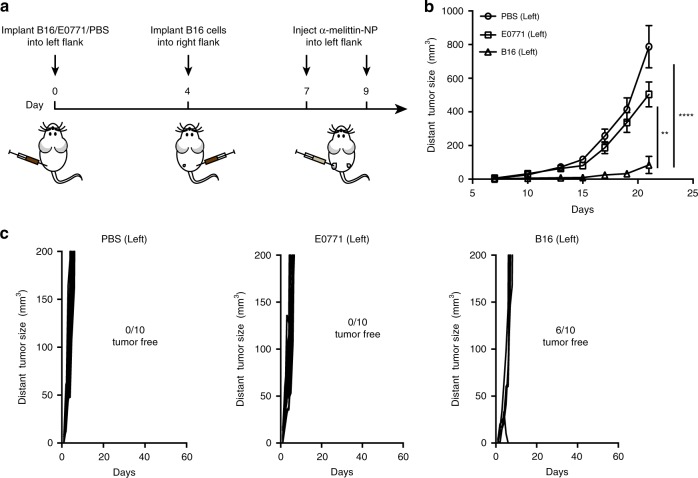
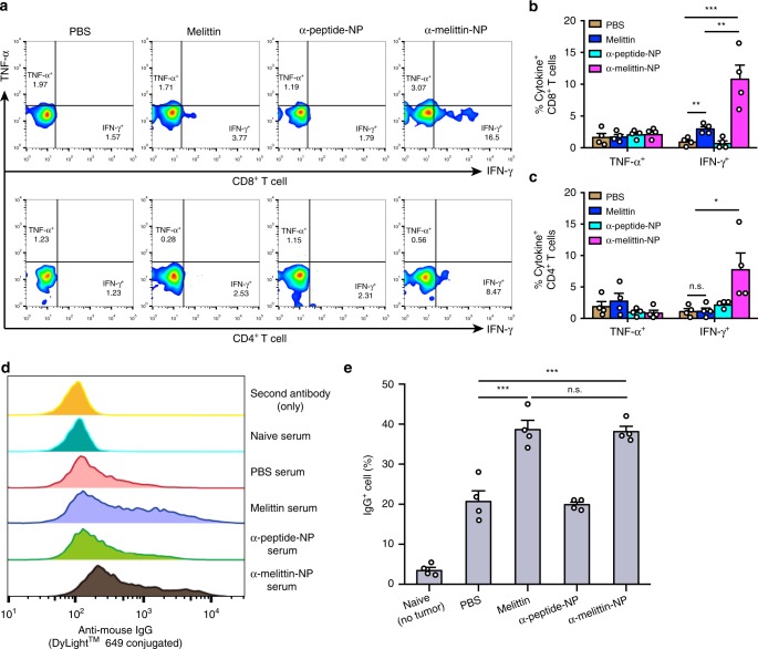
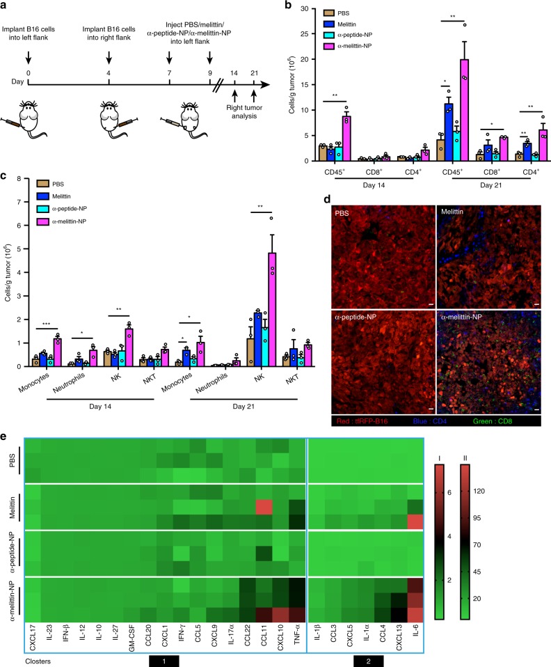
Targeted delivery of a nanovaccine loaded with a tumor antigen and adjuvant to the lymph nodes (LNs) is an attractive approach for improving cancer immunotherapy outcomes. However, the application of this technique is restricted by the paucity of suitable tumor-associated antigens (TAAs) and the sophisticated technology required to identify tumor neoantigens. Here, we demonstrate that a self-assembling melittin-lipid nanoparticle (α-melittin-NP) that is not loaded with extra tumor antigens promotes whole tumor antigen release in situ and results in the activation of antigen-presenting cells (APCs) in LNs. Compared with free melittin, α-melittin-NPs markedly enhance LN accumulation and activation of APCs, leading to a 3.6-fold increase in antigen-specific CD8 T cell responses. Furthermore, in a bilateral flank B16F10 tumor model, primary and distant tumor growth are significantly inhibited by α-melittin-NPs, with an inhibition rate of 95% and 92%, respectively. Thus, α-melittin-NPs induce a systemic anti-tumor response serving as an effective LN-targeted whole-cell nanovaccine.
将负载肿瘤抗原和佐剂的纳米疫苗靶向递送至淋巴结(LNs)是改善癌症免疫治疗效果的一种有吸引力的方法。然而,这种技术的应用受到合适的肿瘤相关抗原(TAAs)的缺乏和识别肿瘤新抗原所需的复杂技术的限制。在这里,我们证明了一种未负载额外肿瘤抗原的自组装蜂毒素脂质纳米颗粒(α-蜂毒素-NP)可促进原位全肿瘤抗原释放,并导致 LNs 中抗原呈递细胞(APC)的激活。与游离蜂毒素相比,α-蜂毒素-NP 显著增加 LN 聚集和 APC 的激活,导致抗原特异性 CD8 T 细胞反应增加 3.6 倍。此外,在双侧侧翼 B16F10 肿瘤模型中,α-蜂毒素-NP 显著抑制原发和远处肿瘤的生长,抑制率分别为 95%和 92%。因此,α-蜂毒素-NP 诱导全身性抗肿瘤反应,可作为有效的 LN 靶向全细胞纳米疫苗。