Hoa Michael, Olszewski Rafal, Li Xiaoyi, Taukulis Ian, Gu Shoujun, DeTorres Alvin, Lopez Ivan A, Linthicum Fred H, Ishiyama Akira, Martin Daniel, Morell Robert J, Kelley Matthew W
Auditory Restoration and Development Program, National Institute on Deafness and Other Communication Disorders, NIH, Bethesda, MD, United States.
National Temporal Bone Laboratory at UCLA, UCLA School of Medicine, Los Angeles, CA, United States.
Front Mol Neurosci. 2020 Feb 5;13:13. doi: 10.3389/fnmol.2020.00013. eCollection 2020.
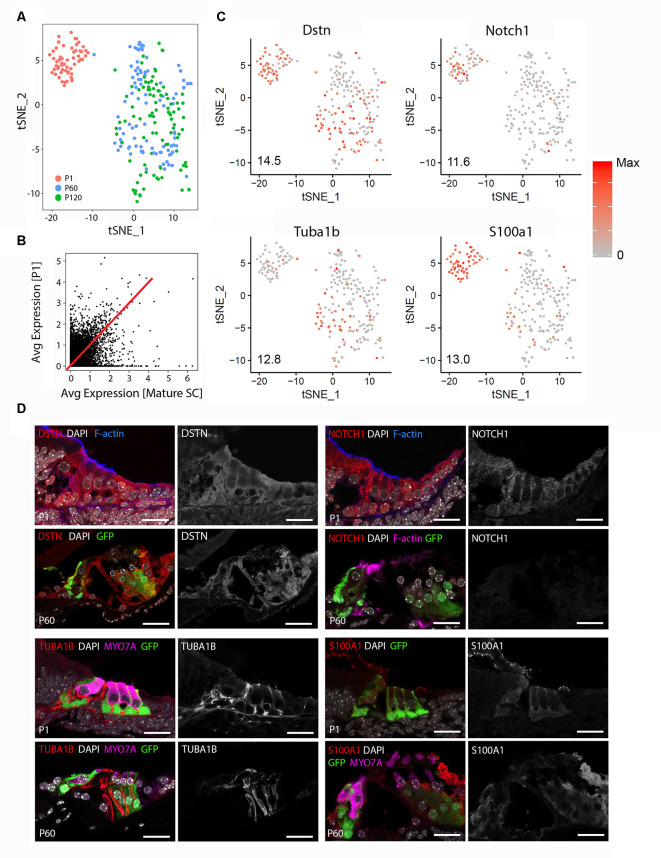
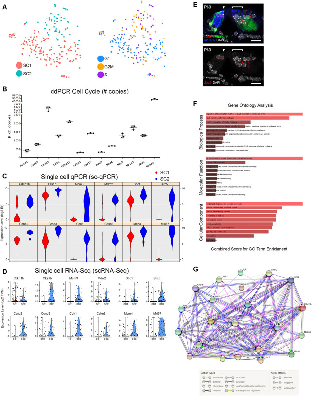
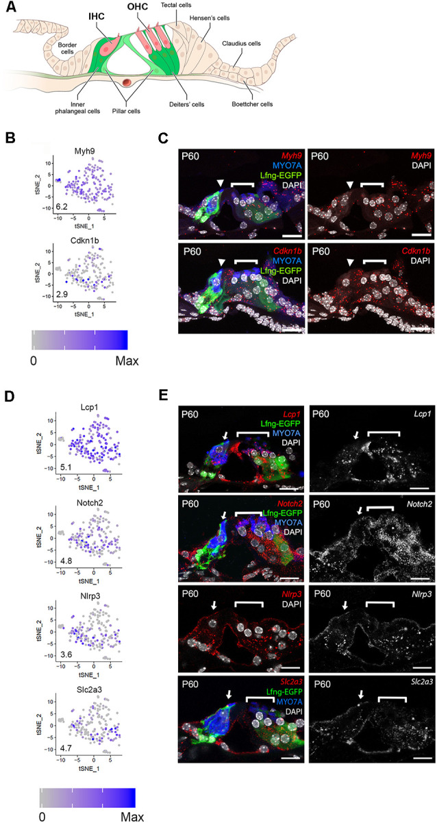
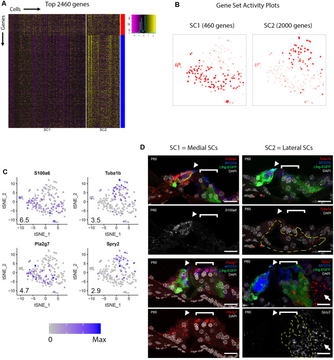
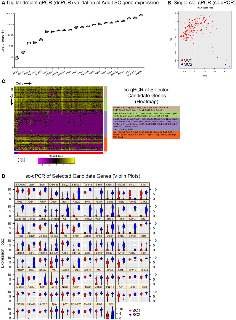
Hearing loss is a problem that impacts a significant proportion of the adult population. Cochlear hair cell (HC) loss due to loud noise, chemotherapy and aging is the major underlying cause. A significant proportion of these individuals are dissatisfied with available treatment options which include hearing aids and cochlear implants. An alternative approach to restore hearing would be to regenerate HCs. Such therapy would require a recapitulation of the complex architecture of the organ of Corti, necessitating regeneration of both mature HCs and supporting cells (SCs). Transcriptional profiles of the mature cell types in the cochlea are necessary to can provide a metric for eventual regeneration therapies. To assist in this effort, we sought to provide the first single-cell characterization of the adult cochlear SC transcriptome. We performed single-cell RNA-Seq on FACS-purified adult cochlear SCs from the adult mouse, in which SCs express GFP. We demonstrate that adult cochlear SCs are transcriptionally distinct from their perinatal counterparts. We establish cell-type-specific adult cochlear SC transcriptome profiles, and we validate these expression profiles through a combination of both fluorescent immunohistochemistry and hybridization co-localization and quantitative polymerase chain reaction (qPCR) of adult cochlear SCs. Furthermore, we demonstrate the relevance of these profiles to the adult human cochlea through immunofluorescent human temporal bone histopathology. Finally, we demonstrate cell cycle regulator expression in adult SCs and perform pathway analyses to identify potential mechanisms for facilitating mitotic regeneration (cell proliferation, differentiation, and eventually regeneration) in the adult mammalian cochlea. Our findings demonstrate the importance of characterizing mature as opposed to perinatal SCs.
听力损失是一个影响相当一部分成年人口的问题。由于噪音、化疗和衰老导致的耳蜗毛细胞(HC)损失是主要的潜在原因。这些人中很大一部分对包括助听器和人工耳蜗在内的现有治疗选择不满意。恢复听力的另一种方法是再生毛细胞。这种疗法需要重现柯蒂氏器的复杂结构,这就需要成熟毛细胞和支持细胞(SCs)都再生。耳蜗中成熟细胞类型的转录谱对于最终的再生疗法而言是必要的,可以提供一个衡量标准。为了助力这一工作,我们试图对成年耳蜗支持细胞转录组进行首次单细胞特征分析。我们对来自成年小鼠的经荧光激活细胞分选(FACS)纯化的成年耳蜗支持细胞进行了单细胞RNA测序,其中支持细胞表达绿色荧光蛋白(GFP)。我们证明成年耳蜗支持细胞在转录上与其围产期对应细胞不同。我们建立了细胞类型特异性的成年耳蜗支持细胞转录组图谱,并通过荧光免疫组织化学、杂交共定位以及成年耳蜗支持细胞的定量聚合酶链反应(qPCR)相结合的方法验证了这些表达图谱。此外,我们通过免疫荧光人颞骨组织病理学证明了这些图谱与成人耳蜗的相关性。最后,我们证明了成年支持细胞中细胞周期调节因子的表达,并进行了通路分析,以确定促进成年哺乳动物耳蜗有丝分裂再生(细胞增殖、分化以及最终再生)的潜在机制。我们的研究结果证明了表征成熟支持细胞而非围产期支持细胞的重要性。